Advertisement

Advertisement

diagnosis

Managing Adolescent Idiopathic Scoliosis (AIS) in Primary Care

Managing Adolescent Idiopathic Scoliosis (AIS) in Primary Care

Teaser: 

Paul J. Moroz, MD, MSc, FRCSC,1 Jessica Romeo, RN (EC), MN, BScN,2Marcel Abouassaly, MD, FRCSC,3

1Children's Hospital of Eastern Ontario, University of Ottawa, Ottawa, Ontario.
2Children's Hospital of Eastern Ontario, University of Ottawa, Ottawa, Ontario.
3Fellow in Pediatric Orthopedic Surgery at the Children's Hospital of Eastern Ontario, Ottawa, Ontario.

CLINICAL TOOLS

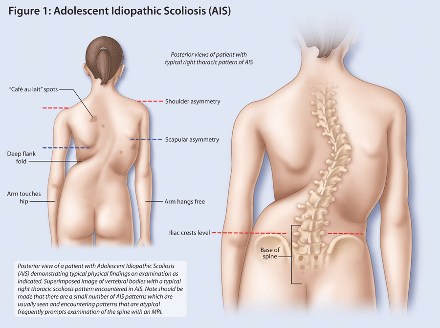
Abstract: Adolescent Idiopathic Scoliosis (AIS) is a condition requiring early detection for appropriate management. Bracing can be effective in preventing curve progression so failing to detect a small AIS curve in a growing child could result in losing the opportunity to avoid a major surgical procedure. Doubts about cost-effectiveness have ended most school screening programs and assessment is now provided mainly by primary care providers. The ability to conduct a quick effective scoliosis examination is important for the busy practitioner. This article illustrates the main features of the screening test, offers guides for imaging, and outlines appropriate tips for specialist referral.
Key Words: Adolescent Idiopathic Scoliosis (AIS), diagnosis, physical exam, Adams Forward Bend Test, primary care.

Members of the College of Family Physicians of Canada may claim MAINPRO-M2 Credits for this unaccredited educational program.

www.cfpc.ca/Mainpro_M2

You can take quizzes without subscribing; however, your results will not be stored. Subscribers will have access to their quiz results for future reference.

1. This can be done with a patient's gown open or closed at the back.
2. The measurement is performed with the examiner sitting and observing the patient from behind. It can be done at the same time as the AFBT, since the examiner is in the same position.
3. With the patient standing erect in bare feet and with the knees extended, the examiner rests his/her hands on top of the iliac crests with fingers extended and palms parallel to the floor. With both the patient's feet flat on the floor, the relative levels of the hands give a surprisingly sensitive estimate of significant LLD (Figure 2).
4. There are alternative methods to measure leg lengths with the patient supine by using a tape measure. These techniques require familiarity with pelvic and ankle landmarks, are time consuming and are remarkably prone to measurement errors.
IMAGING FOR SUSPECTED SPINAL DEFORMITY
1. Radiation exposure using modern radiographic techniques, including digital radiography, is significantly lower than in the past.5
2. Radiologists' reports may use terms related to the spine that can be misleading and worrisome. Cobb angles less than 10 degrees should not be described as scoliosis but rather as "spinal asymmetry" since the term "scoliosis" may prompt an unnecessary referral to a specialist.
3. If imaging is indicated, it is best done at a centre where the patient will be seen in consultation. Radiologists at these centres have the experience to accurately interpret imaging results and correctly report spinal deformity. This also avoids the unfortunate situation where inadequate imaging done elsewhere must be repeated at the referral centre, significantly increasing the patient's radiation dose.
4. Never order a "scoliosis series". It is an obsolete term that referred to pre-operative assessment films. It is still found on some x-ray requisition forms and may be ordered in a misguided attempt to provide the surgeon with as much information as possible. Since the vast majority of patients seen by the spine surgeon will not require surgery, this option is needlessly expensive and the added radiation may be harmful to the patient.
5. The authors allow patients to take smart phone or tablet images of their own spinal x-rays. This engages the patient and their parents or guardians in the management. Take account of all regulatory and privacy issues regarding patient's recording of even their own images.
To have access to full article that these tools were developed for, please subscribe. The cost to subscribe is $80 USD per year and you will gain full access to all the premium content on www.healthplexus.net, an educational portal, that hosts 1000s of clinical reviews, case studies, educational visual aids and more as well as within the mobile app.

Non-Muscle-Invasive Bladder Cancer: Review of Diagnosis and Management

Non-Muscle-Invasive Bladder Cancer: Review of Diagnosis and Management

Teaser: 

Neil Pugashetti,1 Shabbir M.H. Alibhai,3 Stanley A. Yap,1,2

1Department of Urology, University of California, Davis, Sacramento, CA.
2Division of Urology, Department of Surgery, VA Northern California Health Care System, Sacramento, CA, USA.
3Department of Medicine, University of Toronto, Toronto, Ontario, Canada.

CLINICAL TOOLS

Abstract: Non-muscle-invasive bladder cancer (NMIBC) represents the large majority of newly diagnosed bladder tumors and represents a significant burden to both patients and the healthcare system. Although the initial standard treatment for all non-muscle-invasive tumors is surgical resection, there exist a wide variety of both surgical and medical treatment modalities based upon the tumor's specific stage and grade. Ensuring a proper diagnosis is key, and management should be tailored to the individual in order to reduce cancer recurrence and prevent progression of disease.
Key Words: Bladder cancer, non-muscle-invasive, diagnosis, treatment.

Members of the College of Family Physicians of Canada may claim MAINPRO-M2 Credits for this unaccredited educational program.

www.cfpc.ca/Mainpro_M2

You can take quizzes without subscribing; however, your results will not be stored. Subscribers will have access to their quiz results for future reference.

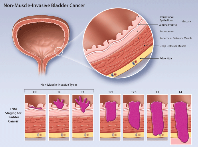
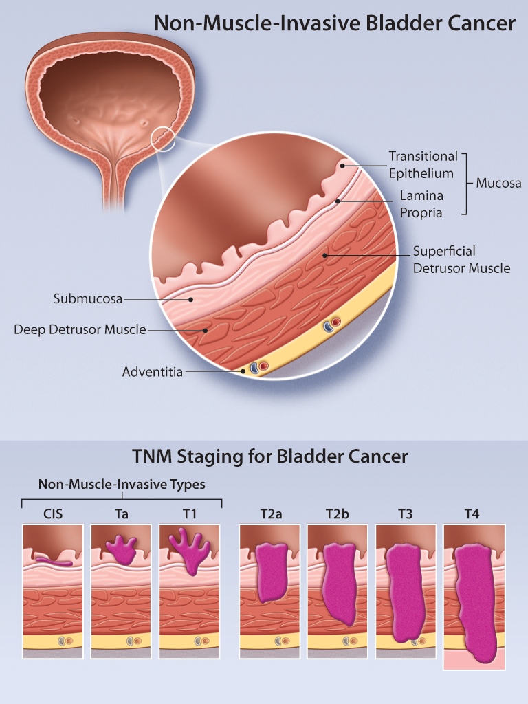
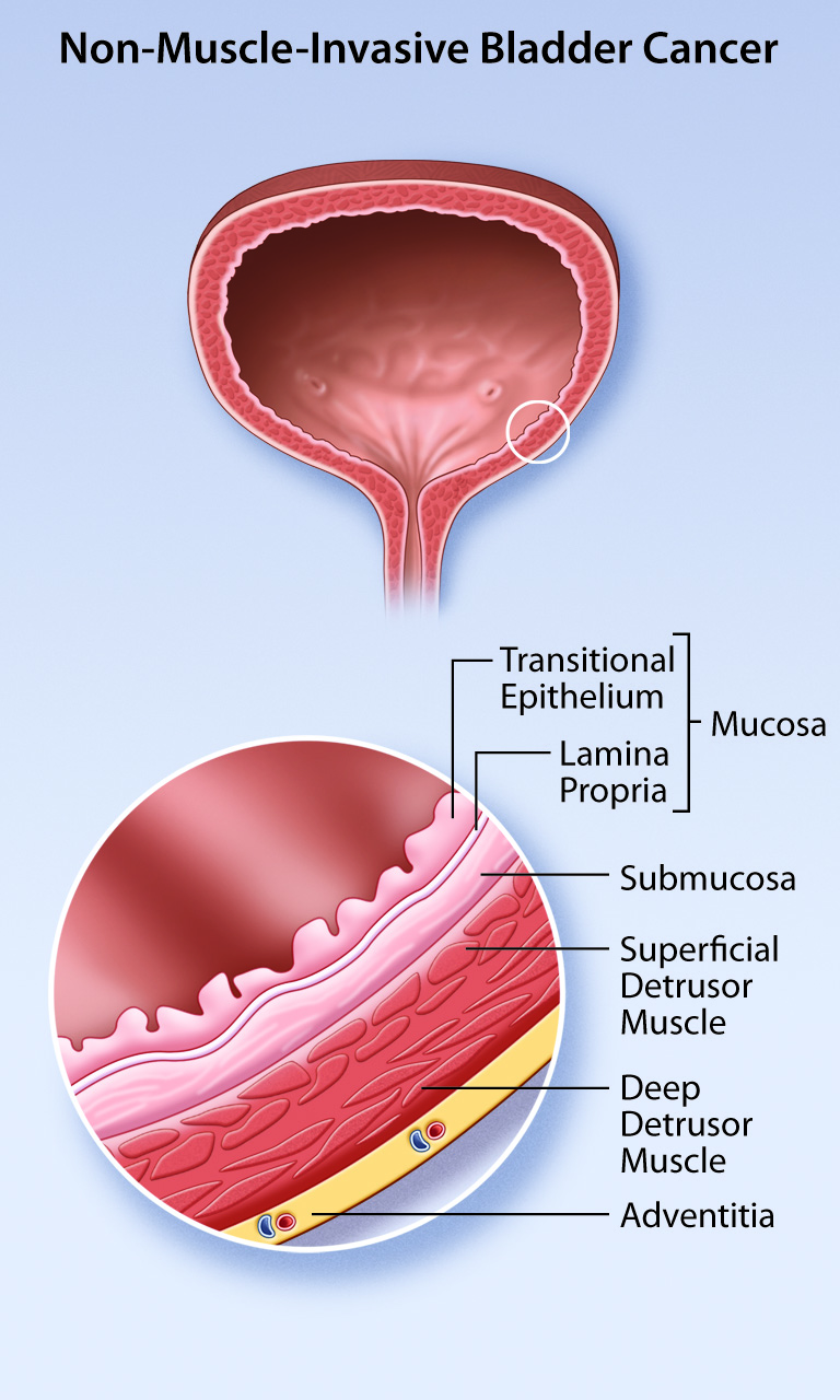
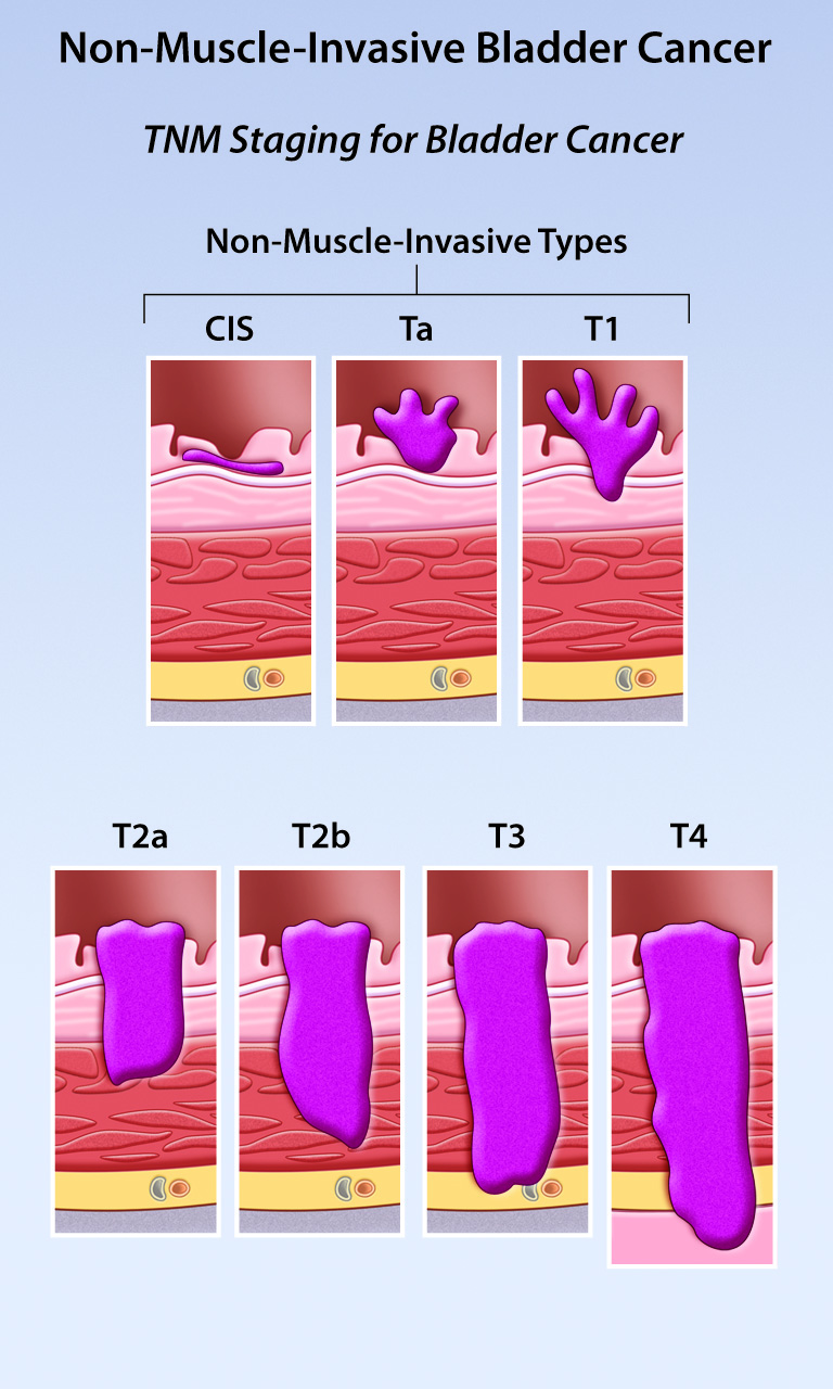
Non-muscle-invasive bladder cancer consists of papillary tumors (Ta), tumors invading the submucosal lamina propria (T1), and flat lesions known as carcinoma in situ (CIS).
Proper management is key given the significant risk of tumor recurrence or progression to muscle-invasive disease.
Many treatment modalities exist including transurethral resection, intravesical chemotherapy, intravesical immunotherapy, and radical cystectomy; treatment choice depends on a variety of factors including tumor stage and grade.
The gold standard for the complete work-up of hematuria is office cystoscopy and imaging of the upper urinary tract.
Initial standard treatment of non-muscle-invasive bladder tumors is TURBT; at the time of resection, sampling of muscle surrounding the lesion is important to accurately assess depth of invasion.
To have access to full article that these tools were developed for, please subscribe. The cost to subscribe is $80 USD per year and you will gain full access to all the premium content on www.healthplexus.net, an educational portal, that hosts 1000s of clinical reviews, case studies, educational visual aids and more as well as within the mobile app.

A Practical Review of the Diagnosis and Management of Small Renal Masses

A Practical Review of the Diagnosis and Management of Small Renal Masses

Teaser: 

Stanley A. Yap,1 Shabbir M.H. Alibhai,2,3Antonio Finelli,1
1Division of Urologic Oncology, Princess Margaret Hospital, University of Toronto, Toronto, ON, Canada. 2Institute of Health Policy, Management, and Evaluation, University of Toronto, Toronto, ON, Canada. 3Department of Medicine, University of Toronto, Toronto, ON, Canada.


Abstract
The incidence of small renal masses (SRMs) has risen steadily over time, and SRMs now represent the majority of newly diagnosed renal lesions. Approximately 80% of newly diagnosed SRMs will be malignant. However, identifying a benign versus malignant lesion non-invasively can be difficult since no distinct imaging characteristics or growth patterns exist between the two. We have witnessed concurrent improvements in treatment strategies for small, localized tumors and have gained a better understanding of their natural history. Along with these changes there has been a shift in the manner in which we diagnose and treat SRMs. Although surgery remains the standard of care, we can now offer a variety of therapies individualized to the patient.
Keywords: kidney cancer, small renal mass, diagnosis, treatment.

Screening for Dementia: First Signs and Symptoms Reported by Family Caregivers

Screening for Dementia: First Signs and Symptoms Reported by Family Caregivers

Teaser: 

Mary A. Corcoran, OTR, PhD, Professor of Clinical Research and Leadership, The George Washington University, School of Medicine and Health Sciences, Washington, DC, USA.

There is an average delay of 20 months between the first recognition of symptoms of Alzheimer’s disease or a related disorder (ADRD) and the seeking of physician help. One reason for this delay is tendency for families to miss early symptoms until the onset of behavioural disturbances. Families may provide more timely accounts with prompted questions. It is important to diagnose cognitive impairment early since there are potential benefits to early treatment. The purpose of this article is to help guide caregivers in identifying a list of symptoms that reflect first indicators of ADRD, based on a study of 68 spouse caregivers of patients with ADRD.
Key words: Alzheimer’s disease, dementia, caregivers, diagnosis, primary care.

After the Fall: The ABCs of Fracture Prevention

After the Fall: The ABCs of Fracture Prevention

Teaser: 

Susan B. Jaglal, PhD, Toronto Rehabilitation Institute Chair, Associate Professor, Faculty of Medicine, Department of Physical Therapy, University of Toronto, Toronto, ON.

A wrist fracture is associated with an increased risk of another fracture and should prompt investigation for osteoporosis in both men and women. If the fracture was caused by low trauma (a fall from a standing height or less), a bone density test should be ordered. If the T score is <–1.5, pharmacological treatment with a bisphosphonate and calcium (1,500 mg/d) and vitamin D3 (≥800 IU/d) is recommended. Management should also include balance, posture, and muscle-strengthening exercises and walking, as well as a review of fall-prevention strategies.
Key words: wrist fracture, osteoporosis, diagnosis, treatment, exercise, falls.

Osteoporosis Screening and Assessment of Fracture Risk

Osteoporosis Screening and Assessment of Fracture Risk

Teaser: 


Mohammed O. Rahman, BHSc student, McMaster University, Hamilton, ON.
Aliya Khan, MD, FRCPC, FACP, FACE, Professor of Clinical Medicine, McMaster University, Hamilton, ON, Director, Calcium Disorders Clinic, St. Joseph’s Healthcare, Hamilton; Director, Oakville Bone Center, Oakville, ON.

Osteoporosis is a skeletal disease characterized by impaired bone strength and an increased risk of fragility fracture. Effective screening should be aimed at evaluating risk factors for osteoporosis with identification of individuals at risk, allowing for intervention prior to fragility fracture. This article presents an overview of the risk factors for fracture in men and women and the integration of these factors in various models, enabling an assessment of the 10-year fracture risk. Through effective screening, early identification, and early intervention with pharmacological therapy of osteoporosis, significant impact can be made on reducing fragility fracture incidence, thereby alleviating the economic and clinical costs to our health care system.
Key words: osteoporosis, screening, risk factors, diagnosis, FRAX.

The Role of the Neurologic Examination in the Diagnosis and Categorization of Dementia

The Role of the Neurologic Examination in the Diagnosis and Categorization of Dementia

Teaser: 

John R. Wherrett, MD, FRCP(C), PhD, Professor Emeritus, Division of Neurology, University of Toronto; consultant in Neurology, Toronto Western Hospital and Toronto Rehabilitation Institute; member, Memory Clinic, Toronto Western Hospital, Toronto, ON.

Nonneurologist practitioners faced with the diagnosis of dementia cannot be expected to conduct the detailed assessments for which neurologists are trained. Nonetheless, they should be able to diagnose the most common forms of neurodegenerative dementia and identify individuals that require more detailed neurologic workup. A neurologic examination algorithm is described that allows the practitioner, in a stepwise and efficient manner, to elicit findings that distinguish the main categories of neurodegenerative and vascular dementia, namely, Alzheimer’s disease, dementia with Lewy bodies, vascular dementia, and frontotemporal lobar degenerations. Patients are assessed for gait, frontal signs, signs of parkinsonism, signs of focal or lateralized lesions, neuro-ophthalmologic signs, and signs characteristic of frontotemporal lobar degeneration.
Key words: neurologic, examination, neurodegenerative, dementia, diagnosis, gait, frontal dysfunction, cognitive impairment.

Chorea among Older Adults

Chorea among Older Adults

Teaser: 

Bhaskar Ghosh, MD, DNB, DM, MNAMS, Movement Disorders Program, Department of Clinical Neurosciences, University of Calgary, Calgary, AB.
Oksana Suchowersky, MD, FRCPC, FCCMG, Movement Disorders Program, Department of Clinical Neurosciences; Department of Medical Genetics, Faculty of Medicine, University of Calgary, Calgary, AB.

Chorea is a hyperkinetic movement disorder characterized by nonsustained, rapid, and random contractions that may affect all body parts. Chorea is hypothesized to be due to an imbalance between the direct and indirect pathways in the basal ganglia circuitry. Important causes of chorea among older adults include medications, stroke, and toxic-metabolic, infective, immune-mediated, and genetic causes. The history and clinical examination guide appropriate investigations and help determine an accurate diagnosis. In secondary causes, removal of the precipitating cause is the mainstay of treatment. If the chorea is persistent or progressive, drug therapy may be instituted. Genetic counselling is important in hereditary chorea.
Key words: movement disorders, chorea, older adults, diagnosis, treatment.

Heart Failure: Old Disease, Older Adults, Fresh Perspective

Heart Failure: Old Disease, Older Adults, Fresh Perspective

Teaser: 


George A. Heckman, MD, MSc, FRCPC, McMaster University, Hamilton General Hospital, Hamilton, ON.
Catherine Demers, MD, MSc, FRCPC, McMaster University, Hamilton, ON.
David B. Hogan, MD, FCRPC, University of Calgary, Calgary, AB.
Robert S. McKelvie, MD, PhD, FRCPC, McMaster University, Hamilton, ON.

The burden of heart failure is rapidly rising. Heart failure is associated with substantial mortality, morbidity, and economic cost, which disproportionately affect older adults. Heart failure among older individuals is frequently complicated by geriatric syndromes, including frailty, functional decline, cognitive impairment, and atypical clinical presentations. Understanding the nature of these geriatric syndromes and their impact on the assessment and management of heart failure is a critical component to diagnosing and delivering appropriate care to these patients. In this article we review the geriatric aspects of heart failure.
Key words: geriatric syndrome, heart failure, older adults, diagnosis, frailty.

Dehydration in Geriatrics

Dehydration in Geriatrics

Teaser: 

MC Faes, MD, MSc, Department of Geriatric Medicine, University Medical Centre Nijmegen, The Netherlands.
MG Spigt, PhD, Department of General Practice/Research Institute CAPHRI, University of Maastricht, The Netherlands.
MGM Olde Rikkert MD, PhD, Department of Geriatric Medicine, University Medical Centre Nijmegen, The Netherlands.

Homeostasis of fluid balance is an important prerequisite for healthy aging. The high prevalence of disturbances of fluid balance among older adult patients has triggered clinical research on age- and disease-related changes in water homeostasis. Empirical findings on risk factors of dehydration and on diagnostic and therapeutic strategies are reviewed in this paper. No single measure has proved to be the gold standard in the diagnosis of dehydration. Diagnosing dehydration and monitoring fluid balance requires repeated measurements of weight, creatinine, and physical signs such as tongue hydration. Rehydration and prevention requires fluid on prescription (> 1.5 litre/day), and the route of fluid administration depends on the acuteness and severity of clinical signs.
Keywords: older adults, dehydration, fluid therapy, risk factors, diagnosis.