Advertisement

Advertisement

Articles

5 Technological Innovations for Those With Dementia

Teaser: 

Holly Clark, Freelance Health Writer

Dementia can be terrifying and devastating to both caregivers as well as the loved ones affected by this disease. People with this disease can now feel some relief due to technological advancement which is meant to improve the quality of their lives. This technology can enhance autonomy as well as independence, manage possible risks in homes and get rid of stress.
In this article, we are going to look at the 5 technological innovations for those with Dementia

1. Communication Aids
Interacting with others is necessary for the quality of life in memory care. Individuals with dementia can remember how an event has made them feel, even if they are unable to recall the faces and names. Technology has simplified the interaction process with loved ones. Adapted telephones are now programmed with contacts that are frequently dialed and usually have bigger buttons which simplify their usage. It's now possible to stay connected with loved ones who are distant apart via the video chat services.
"Changes in the brain caused by dementia begin years before diagnosis. And throughout this timeframe, there are no clear signs that that person has dementia." comments Jane Byrne, Project Coordinator at FirstCare.1

2. Electrical appliance use monitoring

This innovation is meant for caregivers who do not stay together with their loved ones. It controls the use of electrical apparatuses through plugging into a power strip or wall outlet as it will notify caregivers when their appliances have been switched on or off. These technologies do not make the diagnosis of dementia easy. This disease is yet overwhelming. The dementia is now more manageable; this is due to the innovation in new technologies.

3. Reminder messages
Reminders play an important role as the caregiver does as they help to keep the loved ones safe and also retain their relationships. The recording of these messages is done on a device in the residential area and then played out loud at the most suitable time. Caregivers can record a message that when played reminds an individual to take medication at the appropriate time. Some gadgets are designed to play messages based on individuals activity. Some devices are meant to remind individuals with dementia to lock the front door when leaving home. There are other reminder messages designed to help people with dementia on when to close the door when to go to bed and provide reassurance at times when the caregiver is not present.

4. Home care robots Technological advancement has led to the invention of homecare robots which will help reduce the caregiver duty. They are not designed to replace human caregivers, but instead, they are meant to do overall housework and remind individuals who are suffering from dementia on when to take medication or notify medical experts when assistance is required. With further inventions, home care robots may replace caregivers and handle their responsibility fully.

5. In-home cameras
In-home cameras are another technological innovation that is meant to enhance the safety of your loved ones from a distance.2 By either positioning, the camera focusing on medication or in the entrance room can increase your confidence as you are sure your loved one is taking the necessary medication and also active. These cameras can monitor movements and also enable one to communicate with his or her loved one. It will also notify you if no movements have been detected for a particular period.

References

  1. https://www.firstcare.ie/
  2. http://www.scitecheuropa.eu/innovative-technology-dementia/87071/
Disclaimer: 
Disclaimer at the end of each page

Dermatophyte Infections: A Fungus Among Us?

Teaser: 

Miriam Armanious, BSc, MD candidate,1 Joseph M. Lam, MD, FRCPC,2

1Michael G. DeGroote School of Medicine, McMaster University, Hamilton, Ontario.
2Clinical Associate Professor, Department of Pediatrics, Associate Member, Department of Dermatology and Skin Sciences, University of British Columbia, Vancouver, BC.

CLINICAL TOOLS

Abstract: Superficial fungal infections are a common occurrence in adults and children alike. Dermatophytes are the primary cause of these infections, which generally present as erythematous, scaling, annular lesions. Also referred to as "tinea", these infections are classified based on where they are found on the body, as different locations can have slightly different presentations and treatment requirements. This article provides an overview of these various presentations of dermatophyte infections and their risk factors, as well as recommended therapies.
Key Words: dermatophytes, fungal infections, therapies.
Dermatophyte infections, also known as tinea, are very common fungal infections in humans. They occur on the superficial skin, hair, and nails, and can present in many different locations on the body.
Tinea captis is most common in children and can cause hair loss or abscess formation.
When tinea infections are treated with topical corticosteroids, they become harder to detect and are referred to as tinea incognito.
Tinea infections are common, but should be confirmed with KOH microscopy and/or culture from a skin scraping, nail clipping, or hair sample.
Tinea capitis can be mistaken for eczema or seborrheic dermatitis
Check patients who have tinea infection for tinea pedis, since this is a common source of infection for sites on the rest of the body
Treatment for dermatophyte infections can include oral antifungal agents such as terbinafine or grise-ofulvin in a weightdependent dose, or topical antifungal agents. Systemic agents are generally re-served for presentations that penetrate hair follicles and nails, or those that are refractory to topical treatment.
To have access to full article that these tools were developed for, please subscribe. The cost to subscribe is $80 USD per year and you will gain full access to all the premium content on www.healthplexus.net, an educational portal, that hosts 1000s of clinical reviews, case studies, educational visual aids and more as well as within the mobile app.
Disclaimer: 
Disclaimer at the end of each page

A Case of Calcified Nodule in the Base of the Tongue

Teaser: 

Dr. Pradeep Shenoy, MD, FRCS, FACS, DLO,1 Dr. Lyew Warren, MD, FRCPC,2

1ENT & Neck Surgeon, Campbellton Regional Hospital, Campbellton, New Brunswick, Canada.
2Consultant Pathologist, Campbellton Regional Hospital, New Brunswick, Canada.

CLINICAL TOOLS

Abstract: Calcified nodules are uncommon in the base of tongue. Solitary amyloid metaplasia can occur at the base of the tongue and can produce calcification and are difficult to diagnose by histopathological examination.1 Ossoeus choristoma of the tongue is a rare benign condition producing calcified nodule in the base of the tongue.2 Unusual ectopic osseous tongue masses are reported in dermoid cysts or teratomas.3 Osteoma of the base is also reported in the literature.4 Venous malformations with phleboliths are also described in the literature5 causing calcified nodules at the base of tongue.
Key Words: calcified nodules, amyloid metaplasia, osseous choriostoma, dermoid cyst, teratoma and phlebolith.
There are various conditions like ectopic thyroid, lipoma, lymphoma, lymphoid hyperplasia and metastatic lymphatic spread can present with base of tonge swelling. Only a few conditions discussed in the article can have calcified nodule at the base of the tongue.
Base of tongue pathology can be easily missed unless prompt examination, investigations and management is done.
To have access to full article that these tools were developed for, please subscribe. The cost to subscribe is $80 USD per year and you will gain full access to all the premium content on www.healthplexus.net, an educational portal, that hosts 1000s of clinical reviews, case studies, educational visual aids and more as well as within the mobile app.
Disclaimer: 
Disclaimer at the end of each page

Ankylosing Spondylitis and Spinal Fractures

Teaser: 

Andrew Kanawati, BSc, MBBS MSc (Hons) UNSW Mast Anat (UNE) FRACS (Orth),1Nicolas Dea, MD, MSc, FRCSC,2Parham Rasoulinejad, BHSc, MD, FRCSC, MSc, 3Christopher S. Bailey, MD, FRCSC, MSc,4

1 Clinical Fellow, London Health Sciences Centre Spine Program, London, ON.
2Spine Surgeon, Clinical Associate Professor of Neurosurgical and Orthopedic Spine Program, Vancouver General Hospital, University of British Columbia, BC.
3 Assistant Professor, Department of Surgery, Division of Orthopaedic Surgery, Schulick School of Medicine and Dentistry, The University of Western Ontario, London, ON.4 Orthopaedic Surgeon, Division of Orthopaedic Surgery, London Health Sciences Center, and Associate Professor, Dept. of Surgery, University of Western Ontario, London, ON.

CLINICAL TOOLS

Abstract: Ankylosing spondylitis is a seronegative spondyloarthropathy associated with HLA-B27. The main site of pathology is the enthesis (site of tendon insertion). The axial skeleton is affected primarily, with the sacroiliac joints initially involved, with the enthesopathy resulting in fibrosis, calcification and fusion of the sacroiliac joints and spine. There is a high incidence of spine fractures in patients with AS, and there is a high rate of missed fractures, therefore advanced imaging in the form of CT and/or MRI is necessary. Due to their highly unstable nature, surgical management of spine fractures in AS is preferable to non-operative care.
Key Words: Ankylosing spondylitis, spondyloarthropathy, sacroiliitis, spine fracture.

Members of the College of Family Physicians of Canada may claim MAINPRO-M2 Credits for this unaccredited educational program.

www.cfpc.ca/Mainpro_M2

You can take quizzes without subscribing; however, your results will not be stored. Subscribers will have access to their quiz results for future reference.

The spine and sacroiliac joints are the primary site of pathology in AS.
The natural history of the disease causes eventual fusion and kyphosis.
Spinal fractures occur at a higher incidence in AS compared to general population.
There is a high rate of missed fractures, and secondary neurologic complications.
Advanced imaging (CT and/or MRI) is mandatory to rule out fracture, because of high false-negative results of plain radiography.
The patient’s kyphosis must be taken into account when applying full spine precautions for suspected fracture.
Patients must not be forced into extension as this may shift an initially non-displaced fractures.
Loss of flexibility and ankylosis of the spinal column results in long lever arms and behavior akin to a long bone, therefore fractures of the spine are highly unstable and usually require surgical stabilization.
To have access to full article that these tools were developed for, please subscribe. The cost to subscribe is $80 USD per year and you will gain full access to all the premium content on www.healthplexus.net, an educational portal, that hosts 1000s of clinical reviews, case studies, educational visual aids and more as well as within the mobile app.
Disclaimer: 
Disclaimer at the end of each page

Yellow and yellow-brown papules and plaques: Differentiating look-alikes in children’s dermatology

Teaser: 

Lauren Schock, BSc, MD Program,1 Joseph M. Lam, MD, FRCPC,2

1Cumming School of Medicine, University of Calgary, Calgary, AB.
2Clinical Associate Professor, Department of Pediatrics, Associate Member, Department of Dermatology and Skin Sciences, University of British Columbia, Vancouver, BC.

CLINICAL TOOLS

Abstract: Yellow-hued papules and plaques in children can be difficult to differentiate as many causes are rare and may not be frequently outside of specialty pediatric dermatology settings. We will review some of the common and concerning yellow-brown papules and plaques found in infants and children and discuss appearance and distribution, pathophysiology, associated findings, and management.
Key Words: dermatology, pediatric, yellow lesions.
Nevus sebaceous typically grow in proportion with patients in early childhood. Excision should be deferred until adolescence to avoid the use of general anesthetic and an informed decision can be made by the child.
Benign cephalic histiocytosis and juvenile xanthogranuloma are both forms of non-Langerhans cell histiocytosis and are benign and self limited.
Consider a diagnosis of tuberous sclerosis in any child presenting with connective tissue nevi, especially if white macules, angiofibroma, or periungual fibroma are also found.
Screen children with necrobiosis lipodica for retinopathy and neuropathy.
Use your hands – rub a suspected lesion of mastocytosis; if urticaria is elicited (a red, itchy, swollen papule or plaque), you have found Darier's sign. Mastocytosis is likely. Be prepared to treat the child with antihistamines if needed.
Juvenile xanthogranulomas are more common under two years of age, and typically appear on the head and neck. Cutaneous xanthomas often occur overlying tendons, or as grouped papules over the extensor surfaces and buttocks.
To have access to full article that these tools were developed for, please subscribe. The cost to subscribe is $80 USD per year and you will gain full access to all the premium content on www.healthplexus.net, an educational portal, that hosts 1000s of clinical reviews, case studies, educational visual aids and more as well as within the mobile app.
Disclaimer: 
Disclaimer at the end of each page

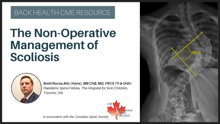
The Role of Screening and Brace Management for Adolescent Idiopathic Scoliosis

Teaser: 

Kedar Padhye, MBBS, DNB (Ortho), 1Reza Ojaghi, MD, 2Fábio Ferri-de-Barros, MD, MSc, FSBOT, FSBOP (Hon.), FRCSC, FCS(ECSA),3

1 Clinical Fellow (Pediatric Spine Surgery)Division of Paediatric Surgery, Section of Orthopaedic Surgery, Alberta Children's Hospital, University of Calgary, Calgary, Alberta.
2Orthopaedic Surgery Resident, Department of Orthopedics, University of Ottawa, Ottawa, Ontario.
3 Department of Orthopedics, Alberta Children's Hospital, Calgary, Alberta.

CLINICAL TOOLS

Abstract: Adolescent Idiopathic Scoliosis (AIS) is defined as curvature of spine in the coronal plane with a Cobb angle of more than 10°. AIS affects 1-3% of children younger than 16 years of age. Less than 20% of those children will progress to severe deformity requiring interventions. Screening with clinical examination and selective radiographic assessment seems to be a cost-effective approach to filter specialist referrals but current literature is controversial. Evidence supports brace management of AIS for skeletally immature patients with primary scoliosis measuring 25°–40. The risk reduction for progression to the surgical range (deformity greater than 50 degrees) is 56%. Timely diagnosis and evidence-based brace management of AIS seem likely to reduce the surgical burden. The implementation of screening guidelines at the primary care level is a critical step.
Key Words: scoliosis; idiopathic; Brace treatment; conservative treatment; screening.

Members of the College of Family Physicians of Canada may claim MAINPRO-M2 Credits for this unaccredited educational program.

www.cfpc.ca/Mainpro_M2

You can take quizzes without subscribing; however, your results will not be stored. Subscribers will have access to their quiz results for future reference.

Bracing is an effective but time sensitive intervention for managing AIS in skeletally immature patients with primary scoliosis measuring 25 to 40 degrees.
Clinical screening is required to identify AIS patients who eligible for bracing.
Improving access to bracing for eligible patients requires a collaborative approach involving primary care physicians and specialists.
1. A systematic collaborative approach involving primary care physicians for screening patients and referring to tertiary care ensures timely assessment and management for eligible patients.
2. Evidence supports brace management of AIS for skeletally immature patients with primary scoliosisl measuring 25°–40°, with the goal of preventing deformity progression to the surgical threshold.
3. A full time (18-23h/day) rigid brace treatment may mitigate the surgical burden of AIS by approximately 30%.
To have access to full article that these tools were developed for, please subscribe. The cost to subscribe is $80 USD per year and you will gain full access to all the premium content on www.healthplexus.net, an educational portal, that hosts 1000s of clinical reviews, case studies, educational visual aids and more as well as within the mobile app.
Disclaimer: 
Disclaimer at the end of each page

Time to Chew on Temporomandibular Disease

Teaser: 

Dr. Robert Caratun, BSc, MSc,1
Dr. Aly Abdulla, MD, CCFP, FCFP, DipSportMed CASEM, CTH, CCPE, McPL,2

1is a graduating medical student from The University of Ottawa going into Family Medicine residency at The University of Calgary in June 2019. He has a background in coaching and creating custom mental skills programs for athletes.
2 is a family doctor with specialties in sports medicine, palliative care, and cosmetic medicine. He can be found on Twitter, LinkedIn and https://ihopeyoufindthishumerusblog.wordpress.com/

CLINICAL TOOLS

Abstract: Temporomandibular disorders (TMD) are one of the most common non tooth-related chronic orofacial pain conditions that involve the muscles of mastication and/or the temporomandibular joint (TMJ) and associated structures. This article reviews the etiology, diagnosis, and treatment of this chronic pain condition.
Key Words: chronic pain, temporomandibular disorders (TMD), temporomandibular joint (TMJ).
1. The etiology of TMD is multifactorial in nature
2. TMD is a clinical diagnosis. Clinicians should perform a complete history and physical with special focus on a dental and psychiatric history.
3. Imaging can be considered if history and physical are insufficient for diagnosis. Diagnostic injections can also be used to further guide clinicians.
4. For TMD treatment, supportive patient education should be prioritized (jaw rest, soft diet, passive stretching) in addition to conservative treatment measures (e.g. NSAIDs).
The most common presenting symptoms of TMD are facial pain, ear discomfort, headache and jaw discomfort/dysfunction.
Symptoms of TMD are typically associated with jaw movement and pain in the temple, masseter, or preauricular region. If there is no pain with jaw movement, consider an alternate diagnosis.
A large volume of patients report abnormal jaw sounds with no jaw pain or dysfunction. Do not treat adventitious jaw sounds; only pain or discomfort in TMD
Patent supportive measures and conservative treatment result in significant pain reduction for the majority of patients and should be the main focus of TMD treatment.
To have access to full article that these tools were developed for, please subscribe. The cost to subscribe is $80 USD per year and you will gain full access to all the premium content on www.healthplexus.net, an educational portal, that hosts 1000s of clinical reviews, case studies, educational visual aids and more as well as within the mobile app.
Disclaimer: 
Disclaimer at the end of each page

Groundbreaking New Study On Ultra-Processed Foods Provides Possible Causal Smoking Gun For Our Global Obesity Struggles

Teaser: 

Yoni Freedhoff, MD,

Family doc, Associate Professor at the University of Ottawa, Author of The Diet Fix, and founder of Ottawa's non-surgical Bariatric Medical Institute—a multi-disciplinary, ethical, evidence-based nutrition and weight management centre. Nowadays I'm more likely to stop drugs than start them. You can also find me on Twitter and Facebook.

CLINICAL TOOLS

Abstract: The reason why weights rise in the industrialized world remains unclear, but most agree that diet plays a crucial role. The endless list of fad diets from paleo to keto to low-carb has led to public mistrust and confusion. The results of a new study titled "Ultra-processed diets cause excess calorie intake and weight gain: A one-month inpatient randomized controlled trial of ad libitum food intake" strongly suggests that regardless of diet, ultra-processed foods should be avoided.
Key Words: diet, nutrition, ultra-processed foods, calorie intake, weight gain.
Helping patients understand the importance of limiting the consumption of ultra-processed foods may be the first step to a healthier lifestyle.
Suggest planning meals ahead of time and eating healthy fats (olive oils, avocado, nuts), grains, enough protein (fish, beans, nuts) and fresh fruit and vegetables.
Discuss how ultra-processed foods may well be a contributor to both weight and other diet related diseases.
Reducing or eliminating consumption of ultra-processed foods may be an effective strategy for obesity prevention and treatment, but doing so requires privilege, time, skill, and expense.
To have access to full article that these tools were developed for, please subscribe. The cost to subscribe is $80 USD per year and you will gain full access to all the premium content on www.healthplexus.net, an educational portal, that hosts 1000s of clinical reviews, case studies, educational visual aids and more as well as within the mobile app.
Disclaimer: 
Disclaimer at the end of each page

5 Ways Technology Is Leading The Revolution In Patient Care

Teaser: 
Rahul Varshneya is the co-founder and President of Arkenea and Benchpoint. Rahul has been featured as a business technology thought leader in numerous media channels such as Bloomberg TV, Forbes, HuffPost, Inc, among others.

Technology is proving itself to be a driving force behind innovations in the healthcare industry. Advances in medical technology are empowering both patients and healthcare providers to take data driven decisions for better health outcomes and improved care efficiency.

Healthcare currently accounts for 17.8 percent of annual GDP spending in the US, which is projected to rise to 19.9 percent by 2025.1 Adoption of technology is slated to play a major role in this growth in healthcare and prove immensely beneficial for everyone within the chain of care.

Here are the ways technology is leading the revolution in patient care:

1. Rise in Electronic Health Records
Data is the backbone of technological developments and widespread adoption of EHRs across hospitals has made the collection and analysis of healthcare data an easy task. 86.9 percent of all physicians make use of EHRs to store patient data.2

Storage, maintenance and analysis of this data is important for efficient monitoring of the patients. Advances in computing methods, big data analytics and use of artificial intelligence to sift through medical data at revolutionary pace and obtain meaningful results is all contributing towards better patient outcomes.

Adoption of EHRs comes with it's own set of challenges, data security being the biggest threat of all. According to research, healthcare industry is subjected to 340 percent more security incidents than any other industry and is 200 percent more likely to encounter data theft.3 Storage of medical data thus has to be done ensuring the security measures are in place and sensitive patient data is always protected.

2. Increased Adoption of Telehealth
Telehealth is revolutionizing patient care by making healthcare services more accessible to all. CMS recently proposed it's 2019 Medicare Physician fee Schedule and Quality Payment Program that would result in increased adoption of telehealth services.4 The proposed changes would result in advancing virtual care for patients by leveraging technology, laying down norms for physician reimbursement for telehealth services, thus reducing physician burden.

The widespread use of smartphones and advances in mobile networks and connectivity has enabled the physicians to have virtual consultations with the patients. It negates the need for the patient to travel down to the physician's office for a routine health consultation which is extremely important in case of chronically ill and debilitated patients.

It also makes patient management more efficient by streamlining patient appointments and reducing wait times. Remote patient monitoring results in better health outcomes while reducing healthcare costs. Use of telemedicine to tackle ER triage recorded a 25 percent reduction in staffing costs in the hospital, while increasing the admission rate by 20 percent.5

The telehealth market is already growing fast and is projected to reach 52.89 billion by the end of 2025.6 It has already transformed patient care and further technological advancements like advent of 5G technology will give it a further boost in the days to come.

3. Wearable Tech and Internet of Medical Things
One of the most important technological revolutions in recent times has been the advent of wearable devices. The smartwatches sale is projected to reach 86 million units by 2021 which will be only 16 percent of all wearable devices.7

Equipped with state of the art sensors, these fitness and medical devices track the individual health stats empowering it's wearer to take conscious, data driven health decisions.

The healthcare data collected by these devices is also utilized by the healthcare providers in order to curate customized care plans on the basis of individual needs. The in-built sensors detect any abnormality in the readings resulting in an early diagnosis of the underlying conditions.

Use of Artificial intelligence tools to compute the data collected by these devices can help predict disease trends across populations and bring about a data driven revolution in the field of medical research.

4. Patient and Workflow Management
Leveraging technological tools like Artificial Intelligence8 to automate the routine tasks in patient management can ensure that the doctors and nurses9 can prioritize on the more important tasks on their hands. Use of technology to manage things like patient registration, filling in the notes in patient's medical records, processing discharge and payments not only results in saving time and resources within the hospital, but it also makes the workflow within the hospital more organized and optimized.

Use of self serving kiosks for patient registry, voice to text input of patient data into medical records, use of chatbots for routine conversations with patients to ensure patient compliance are some of the ways in which technology is transforming patient management.

Automation of the routine non-emergency tasks would result in better focus on the emergent cases and more time spent by both doctors and nurses at the patient's bedside, resulting in greater patient satisfaction and improved patient outcomes. An optimized hospital workflow also results in optimal usage of resources thus saving operational costs.

5. Mobility in Healthcare

Mobility in Healthcare10 is going to undergo a revolutionary growth of 28.3 percent by 2022 allowing the focus to shift from hospital based care to a more patient centric approach.11

Development of robust mobile apps12 empowers the physicians to provide the best possible treatments, resulting in more positive patient outcomes and overall decrease in treatment costs. Rise of mobility has made it possible for the patient data from the wearable devices to be integrated into the EHRs resulting in a more holistic care plans to be designed for specific patients.

Mobility in healthcare also results in better workflow optimization within the hospital by tracking the real time location of the healthcare providers resulting in better access and communication.

Summing up
The field of healthcare has historically been one of the last segments to adapt to rapidly changing technology. The scenario is now transforming with the gradual inflow of advancements which have resulted in a renovation of the healthcare sector. While the applications are plenty and the transformation has just begun, one thing is for sure, incorporation of technology in healthcare is leading us towards a brighter future.

References

  1. https://www.cms.gov/newsroom/press-releases/2016-2025-projections-national-health-expenditures-data-released
  2. https://www.cdc.gov/nchs/data/factsheets/factsheet_nhcs.pdf
  3. www.forcepoint.com/content/2015-industry-drill-down-report?utm_source=Websense&utm_medium=Redirect&utm_content=2015-finance-industry-drilldown%3
  4. https://www.cms.gov/newsroom/press-releases/cms-proposes-historic-changes-modernize-medicare-and-restore-doctor-patient-relationship
  5. https://mhealthintelligence.com/news/hospitals-turn-to-telemedicine-to-tackle-er-triage-overcrowding
  6. https://www.prnewswire.com/news-releases/telehealth-market-to-2025--global-analysis-and-forecasts-300705902.html
  7. https://www.ccsinsight.com/press/company-news/2968-ccs-insight-forecast-reveals-steady-growth-in-smartwatch-market
  8. https://www.entrepreneur.com/article/325436
  9. https://myresumeseed.com/nurse-practitioner-resume/
  10. https://www.mgma.com/resources/health-information-technology/healthcare-mobility-trends-with-greatest-potential
  11. https://www.marketresearchfuture.com/reports/healthcare-mobility-solutions-market-1970
  12. https://arkenea.com/mobile-app-development/
Disclaimer: 
Disclaimer at the end of each page

Pregnancy-Related Back Pain: When Should I Worry?

Teaser: 

Dr. Julia Alleyne, BHSc(PT), MD, CCFP, Dip. Sport Med MScCH,

is a Family Physician practising Sport and Exercise Medicine at the Toronto Rehabilitation Institute, University Health Network. She is appointed at the University of Toronto, Department of Family and Community Medicine as an Associate Clinical Professor.

CLINICAL TOOLS

Abstract: Clinicians are often unsure if back pain during pregnancy is due to a musculoskeletal condition, an abnormality with the pregnancy or merely part of the common discomforts associated with gestational changes. Low back pain guidelines do not include pregnant women in their criteria and there have been no randomized clinical trials to determine specific causes of low back pain during pregnancy. This article will provide the clinician with a framework for identifying pregnancy-related back pain using a high yield history and key physical examination techniques to differentiate between mechanical back pain, sacroiliac instability and symphysis pubis separation. Risk factors for low back pain and warning signs for pregnancy complications will be identified. Appropriate management strategies will be provided for the management of pregnancy-related low back pain.
Key Words: pregnancy-related low back pain, pregnancy, pelvic pain, physical examination, management.

Members of the College of Family Physicians of Canada may claim MAINPRO-M2 Credits for this unaccredited educational program.

www.cfpc.ca/Mainpro_M2

You can take quizzes without subscribing; however, your results will not be stored. Subscribers will have access to their quiz results for future reference.

1. Probable Risk Factors for Low Back pain during pregnancy include: • Pre-pregnancy and past pregnancy low back pain • Low Back and Pelvic Trauma • Poor general physical condition • Joint Hypermobility • Increase body weight
2. Pregnancy related low back or pelvic pain is defined as intermittent or constant pain in the lumbar, buttock, pelvis, groin and/or upper thigh area lasting for more than one week.
3. Exercise, education and postural advice are the mainstays of treatment and can be enhanced by short term therapy with a rehabilitation professional.
1. Patients who have low back pain, in any trimester, associated with vaginal bleeding, uterine contractions, fever or hematuria should be immediately referred for obstetrical consultation.
2. The three most common causes of low back pain in pregnancy are mechanical low back strain, sacroiliac instability and symphysis pubis separation; they often occur together.1,2
3. Patient with Symphysis Pubis Dysfunction complain of significant pain during most of these activities: • Walking • Climbing Stairs • Turning in Bed • Standing on one Leg • Rising from a Chair
To have access to full article that these tools were developed for, please subscribe. The cost to subscribe is $80 USD per year and you will gain full access to all the premium content on www.healthplexus.net, an educational portal, that hosts 1000s of clinical reviews, case studies, educational visual aids and more as well as within the mobile app.
Disclaimer: 
Disclaimer at the end of each page