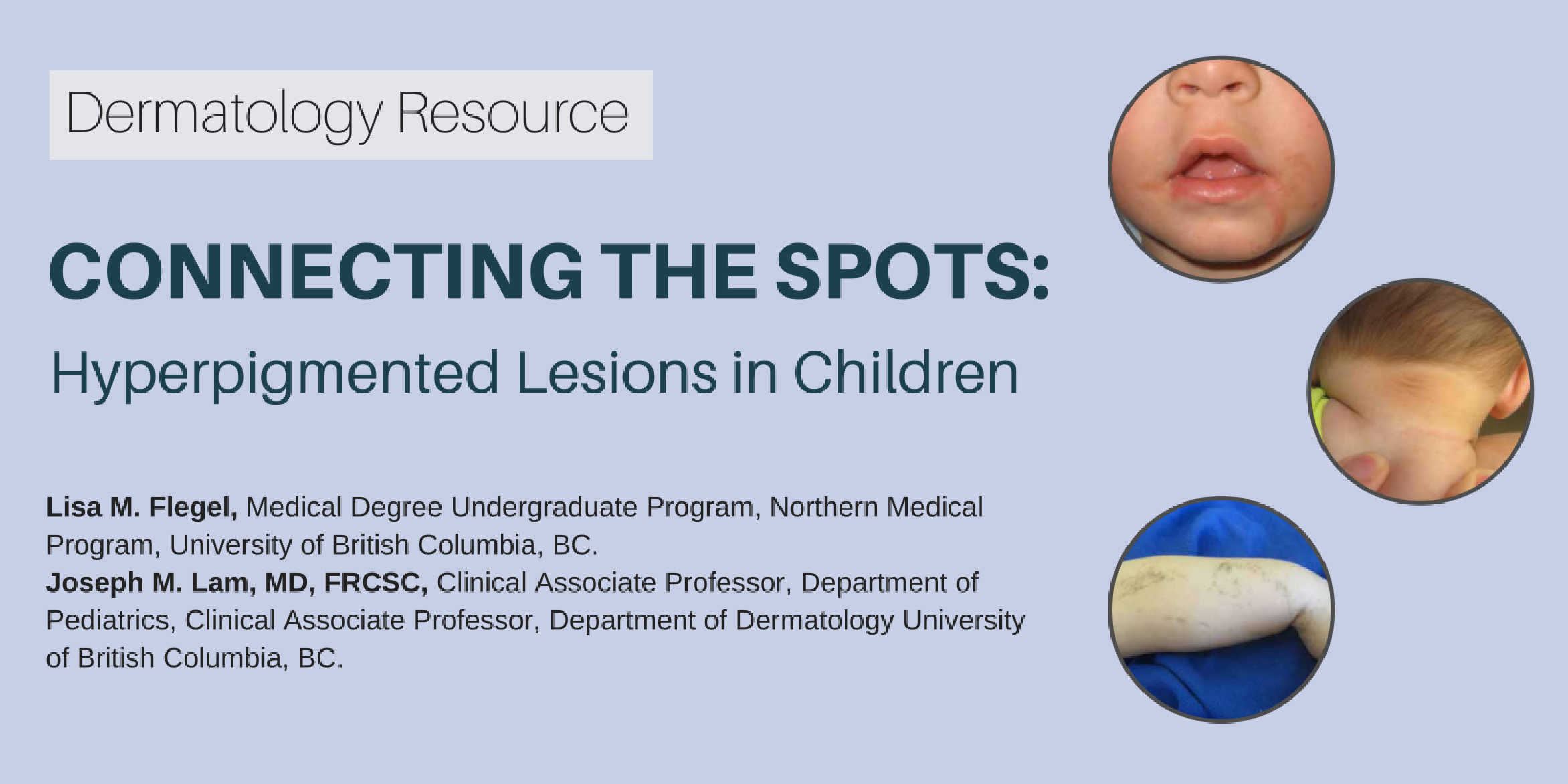
Connecting the Spots: Hyperpigmented Lesions in Children
| Questions | 5 |
|---|---|
| Attempts allowed | Unlimited |
| Available | Always |
| Pass rate | 75 % |
| Backwards navigation | Allowed |
| Questions | 3 |
|---|---|
| Attempts allowed | Unlimited |
| Available | Always |
| Pass rate | 75 % |
| Backwards navigation | Allowed |
| Questions | 5 |
|---|---|
| Attempts allowed | Unlimited |
| Available | Always |
| Pass rate | 75 % |
| Backwards navigation | Allowed |
| Questions | 3 |
|---|---|
| Attempts allowed | Unlimited |
| Available | Always |
| Pass rate | 75 % |
| Backwards navigation | Allowed |
One always hopes that as medical practitioners, we will be able to focus our attention on the medical issues faced by seniors and help families cope with the fears, disappointments and tragedies that are faced by loved ones in the midst of what are often life-altering illnesses.
Throughout our initial medical training, and most often during post-graduate training programs, the primary focus in general is: what is the "best of medicine" and what does "evidence-based medicine" tell us about treatment decisions and their ultimate impact on health, well-being and, often, the likelihood of death? This is particularly the case in the care of the older adult—whether in geriatric medicine or eldercare.
What is often surprising and baffling, especially to younger physicians, is the situation where the core of what appears to be the challenge in care provision is negatively tinged by what might be called family "strife." At times, however, a more appropriate term would be venomous, hateful actions—actions that ultimately will be destructive to the family fabric.
This should not be surprising to anyone who has even a modest understanding and familiarity with the world of literature—whether limited to English works, or more broadly including European or other literature.
Those medical trainees who have worked with me have in all likelihood heard me either seriously or humorously say, "If I were king, all first degrees would be in English literature." Or when there is a complex family dynamic playing out, I might say, "It's King Lear—if you have not read it ever or lately, read it or read it again—it's all there."
Sometimes I feel like that great American comic Jimmy Durante, who was quoted as saying, "I have a million of them, a million of them," referring to his often delectable jokes. According to an online biographical history, it has been said that "I've got a million of 'em" is what Durante (1893-1980) often said after telling a corny joke. Durante was credited with "I've got a million of 'em" in a 1929 newspaper story.
I say this when referring to complex family situations in which what appears to be the worst in human interactions seems to be playing out. Often the issue is related to money (or property), and if one is in a position to hear the story from all the parties, it often becomes clear that, for whatever reason, the pot has come to a boil at this juncture of life. This is usually because the flame heating the water that's not boiling has been on for what appears to have been many years.
Most of us know of such stories, hopefully not in our own families, but it is unlikely that there is a family who is not familiar with a "Lear-like" scenario in someone close to them. Greed, jealousy, hurtful memories, mean-spirited personalities, events that occurred—sometimes decades earlier—that were never resolved or left indelible scars are often the reasons cited for the enmity.
I have had the good fortune to observe that, on some occasions, especially when a parent, in particular, is dying, though it could be another relative, there is the possibility of repairing longheld animosities and bringing long-estranged family members back together. It does not always succeed, but I have witnessed the monumental efforts of health-care staff—especially those in social work, nursing and medicine, although any and all of the health-care staff can be key—in bridging the emotional moat that often separates family members.
It may not always work, but I believe it is always worth the effort. Living with the result of lifelong family strife is often disabling, and the scars that occur and that are left can have long-lasting negative effects on people's lives and their own abilities to have meaningful and binding relationships with their siblings and offspring.
This article was originally published online at http://www.cjnews.com/perspectives/opinions/dealing-family-strife
D’Arcy Little, MD, CCFP, FRCPC
Medical Director, JCCC and HealthPlexus.NET

Physicians usually become adept at choosing medications for the complaints and illnesses that patients bring to their attention. Doctors have to become familiar with the common medications that are indicated for the most prevalent illnesses they see, and there are many resources available to keep physicians as up to date as possible on the most effective drugs and what the medical evidence says about indications, side effects, drug interactions and priorities of care at various points in the progression of a patient’s illness. In the elderly, there are often a number of illnesses competing for possible medical attention and intervention.
Dementia is the umbrella term used commonly to describe the cognitive decline that affects many older individuals. It may be due to a number of recognized conditions of which Alzheimer’s is the most commonly recognized, but the effects of blood vessel (vascular) disease are also very common factors in the aging population.
There are some medications for these conditions that affect memory, judgment and behaviour, the symptoms of which may cause great strife in the individuals affected as well as their families. The symptoms often cause immense challenges when it comes to the use of possibly helpful medications. The pharmaceutical products available for improvement of memory and judgment are often helpful in some individuals, but even when they are effective, they do not “cure” the cognitive impairment. They may, however, provide some improvement in certain aspects of cognition and especially socialization and interactive abilities.
Most challenging are the medications available for what are called behavioural manifestations of dementia, so much so that decisions to transfer to protective living environments such as nursing homes may be the result of such behavioural processes. These events may occur periodically and in what appear to be unpredictable outbursts. Although there are medications that are often used under such circumstances, which may be effective in decreasing the intensity of the disturbing symptoms, they—as do all medications—have potentially bothersome side-effects that may limit their efficacy.
During the past few years, the medical and non-medical health-care professionals involved in such care decisions have discovered that a number of non-medication interventions may be very effective and helpful without the risk of medication side-effects. Probably the most well-acknowledged and studied has been the use of individualized music, which has been shown to quell some of the agitations and disruptive behaviour associated with dementia. There are programs through the Alzheimer societies that provide personalized music on small iPods that can be used during episodes of behavioural outbursts.
In addition, there has been an expanding experience of using a range of alternative treatments such as pet therapy and doll therapy. In the latter, agitation, primarily in women, can be calmed by providing a life-like infant doll that brings out the calming and nurturing reactions many older women experienced during their earlier maternal days. Massage therapy and aroma therapy have also been used with good results in certain individuals.
The importance of these alternative therapies is that, unlike medications, they usually do not have side effects that might limit their effectiveness. They often tap into aspects of the person’s residual abilities that bring out what might otherwise be hidden aspects of his or her personality. Of greatest benefit is that these therapies are often provided by concerned and loving family members or dedicated health-care professionals, thus enhancing the social aspects of care that have been identified as being important through the course of conditions responsible for cognitive impairment.
Just imagine listening to one’s favourite music with an affectionate cat on one’s lap, while someone who cares enough provides a hand massage, rather than a dose of a medication that may cause drowsiness, increase the risk of falls and impair the person’s ability to walk securely. It may not always work, but it is always worth a try. So it’s important to be persistent and see what might work.
This article was originally published online at http://www.cjnews.com/perspectives/opinions/beyond-medications-dementia