Advertisement

Advertisement

dementia

Antiamyloid Immunotherapy: What Do the Blood Vessels Think?

Antiamyloid Immunotherapy: What Do the Blood Vessels Think?

Teaser: 

Click here to view the entire report from the 4th Canadian Colloquium on Dementia

Antiamyloid Immunotherapy: What Do the Blood Vessels Think?

Speaker: Steven M. Greenberg, MD, PhD, Director, Hemorrhagic Stroke Research Program, Massachusetts General Hospital; Associate Professor of Neurology, Harvard Medical School, Boston, MA, USA.

The final speaker, Dr. Steven Greenberg, focused on vascular issues related to antiamyloid immunotherapies.

Is Antiamyloid Therapy the Solution?
He began by addressing whether antiamyloid therapy might be considered a solution or might only contribute further to the problem of cognitive deterioration, as well as vascular pathology. As Dr. Relkin had discussed, earlier significant trials of the therapy resulted in a serious inflammatory response. What possible approaches might avoid this toxicity?

Historically, Dr. Greenberg observed, people taking anti-inflammatories to retard inflammation were thought to be at decreased risk for Alzheimer’s disease (AD), as inflammation was perceived as part of the cascade leading to AD-associated deterioration. That was turned on its ear in Dr. Dale Schenk’s work, as discussed by Dr. Relkin. Schenk’s work on immunization to beta-amyloid with mouse models appeared to generalize to humans. Indeed, individuals who have participated in immunization trials and who came to autopsy showed striking clearing of beta-amyloid deposition as a result of the therapy. The serious drawback of this treatment was the risk of meningoencephalitis, which led to initial discontinuation of the study.

The Toxicity of Antiamyloid Therapy
The adverse events included subacute cognitive decline, seizures, and white matter changes. These effects were reversible in some cases; however, two deaths were reported. The value of passive immunotherapy is still emerging-studies in which patients are receiving premade antiamyloid antibody therapies are ongoing, and some changes, reminiscent of the previous white matter changes on MRI, have been reported. Passive immunotherapies may not totally avoid toxicity and severe side effects.

Cerebral Amyloid Angiopathy and Inflammation
Dr. Greenberg stressed the connection between meningoencephalitis and cerebrovascular amyloid or cerebral amyloid angiopathy (CAA). This topic partners with the study of AD and amyloid. It involves essentially the same peptide, with a difference in Ab40 and Ab42 ratios. Dr. Greenberg showed an illustration of the life cycle of the amyloid peptide, a complex entity (Figure 1). Once formed, it might degrade, be deposited in the vessels, or pass out of the nervous system. There is a strong pathological overlap between Alzheimer’s and CAA. Research has uncovered the prevalence of vascular amyloid in almost every AD case. Dr. Greenberg also emphasized the presence of microbleeds which are, radiographically, a hallmark pathological feature found in a substantial set of AD patients. Such hemorrhages are also a cardinal feature of amyloid angiopathy, which tends to be worse in the parietal lobes where the hemorrhages are located. There is a nearly superimposable distribution of the hemorrhages when one examines individuals with AD; the microbleeds in AD patients occur in the same distribution as amyloid angiopathy. Bleeding in AD patients, Dr. Greenberg contended, suggests underlying amyloid angiopathy. There are virtually no AD patients without this associated vascular pathology.

Dr. Greenberg further considered what this vascular pathology present in AD patients has to do with the toxic response to beta-amyloid immunotherapy. The evidence comes from the pathology of vaccine-associated meningoencephalitis; the resemblance of iatrogenic toxicity to spontaneously occurring syndromes of amyloid angiopathy-related inflammation; and animal studies.
Dr. Greenberg showed brain pathology images from the two fatalities that occurred in the active vaccine study. Both autopsies revealed substantial clearing of amyloid plaques. However, the vascular amyloid remained present at an advanced stage, with evidence of vascular breakdown. There was significant inflammation around the vessels and an outpouring of inflammatory cells, mostly lymphocytes. Pathologically, it looked like there had been an inflammatory response to the vessels themselves. These results, he stated, are striking in light of spontaneously occurring syndromes of CAA-related inflammation: a sizeable subset of patients present with subacute cognitive changes or seizures. The pathology they exhibit is reminiscent of the changes seen in the active vaccine study-including advanced amyloid angiopathy and perivascular inflammation. There are focal cortical infarctions, possibly representing effects of inflammation. Their MRIs show subcortical changes in the white matter, extending to overlying grey matter. Radiographic as well as clinical data suggest similarities between the two processes. Dr. Greenberg compared the amount of white matter disease in study participants with spontaneously occurring CAA syndrome with patients with the iatrogenic pathology from the active vaccine study. Patients treated with anti-inflammatories improved clinically and showed corresponding improvement of white matter findings on MRI. There was a relapsing subgroup with bouts of spontaneously occurring CAA-people who came in with a second or third bout of the same syndrome-who showed repeated instances of return of the hyperintensities.

Studies of transgenic mice with robust plaque and vascular amyloid showed occurrences of hemorrhages in passive immunization. These findings with animal models represent another adverse occurrence and raise the question of what happens to vessels when amyloid is pulled from them.

So, Dr. Greenberg queried, if we suppose that vascular amyloid is the problem and is the trigger of adverse events in immune-based therapy, how is it avoided? Approaches to avoiding CAA-related toxicity might include better noninvasive detection of CAA; better identification of those at risk for CAA-related inflammation; and adjusting the mode of treatment.

Dr. Greenberg considered PET imaging for the noninvasive detection of amyloid angiopathy. He stated that Pittsburgh compound B (PIB) accumulates in regions with high levels of plaques. Because patients with amyloid angiopathy showed increased accumulation of this compound, he questioned whether this signifies that PIB binds to vascular amyloid. Dr. Greenberg argued that PIB predominantly accumulates in the occipital cortex, a region the most highly enriched in vascular amyloid. One may use a ratio of occipital to global PIB binding to determine how much amyloid would be present.

Talking about risk factors, Dr. Greenberg noted the overrepresentation of the APOE 4/4 genotype among patients with inflammatory CAA. This marker may represent susceptibility for CAA-related inflammation.

Finally, altered methods of treatment may minimize the risk of bad effects from vascular amyloid. Dr. Greenberg’s team has worked with multiphoton microscopy to look at the progression of vascular amyloid in living mice and mapped a predictable week-by-week progression of amyloid angiopathy in transgenic mice. The results were measured in a quantitative way across multiple imaging sessions. They wondered what would happen were they to directly apply beta amyloid antibodies to the surface of the brain in these transgenic mice. Results showed a decline in vascular amyloid. The direct application of antibodies to the surface of the brain reversed accumulation to the same extent vascular amyloid would tend to accumulate, with lack of toxicity. He stated that this suggests that beta-amyloid clearance can be done in a way that is protective of the vessels.

Conclusion
Dr. Greenberg concluded that the anti–beta-amyloid inflammatory response may be both beneficial when it is clearing amyloid, but harmful if it is causing tissue damage around it. There are clinical, radiological, animal, and other data suggesting that vascular amyloid contributes to vaccine-related toxicity. The possible approaches may involve altering the therapeutic approach, and determining who should be treated. Selecting subjects at low risk for adverse events may make antiamyloid immunotherapy a safer treatment.

The Pros and Cons of the Amyloid Hypothesis of Alzheimer’s Disease

The Pros and Cons of the Amyloid Hypothesis of Alzheimer’s Disease

Teaser: 

Click here to view the entire report from the 4th Canadian Colloquium on Dementia

The Pros and Cons of the Amyloid Hypothesis of Alzheimer’s Disease

Speaker: Andrea LeBlanc, PhD, Professor and McGill Dawson Scholar, Department of Neurology and Neurosurgery, McGill University; Project Director, Bloomfield Centre for Research
in Aging, Lady Davis Institute for Medical Research, Sir Mortimer B. Davis Jewish General Hospital, Montreal, QC.

Researcher Dr. Andrea LeBlanc addressed evidence for and against the amyloid beta (Ab) hypothesis of Alzheimer’s pathogenesis.

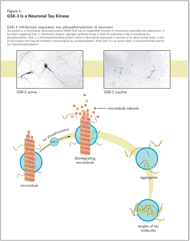
Historically, Alzheimer’s disease (AD) has been characterized by its pathological signs-namely, plaques. But AD pathology encompasses more than amyloid and tau, she stated. There are degenerating neurites within those plaques that are important, as well as neurofibrillary tangles, tau, and so forth. In AD, other features play a role: synaptic loss is obvious and related to dementia but is often not discussed. Also implicated in the degenerative process are glial activation and neuronal loss. The progressive cell loss in AD is not very obvious. So, Dr. LeBlanc questioned, is it neuronal loss, neuronal atrophy and/or neuronal dysfunction causing the cognitive decline?

Arguments Marshalled in Defense of the Amyloid-Beta Hypothesis
The hypothesis holds that the Ab peptide leads to synaptic degeneration, neuronal degeneration and formation of neurofibrillary tangles and thus to the cognitive losses observed. Progressive amyloid deposition is certainly greater in AD patients, Dr. LeBlanc granted. However, some noncognitively impaired individuals have a high plaque burden. Those who defend the hypothesis suggest that they may manufacture a nontoxic form of amyloid. The correlation between amyloid deposition and impairment levels is not clear-cut; for those who doubt the amyloid hypothesis, this finding seems to disprove it.

The second argument often offered by proponents of the Ab hypothesis is that genetic mutations or polymorphisms associated with AD lead to the overproduction of Ab peptide. This is definitely true, Dr. LeBlanc stated-the amyloid precursor protein (APP), presenilin-1 and presenilin-2 genes, and related mutations have been identified by Dr. Peter St.-George Hyslop and colleagues. All of these mutations lead to increased Ab peptide levels, an increase of Ab42 versus Ab40 ratios, or an increase in Ab aggregation in the absence of total increase in Ab. These features do influence the level of Ab or its ability to aggregate. The caveat Dr. LeBlanc added is that the increases are not large-they are statistically significant increases, but their biological significance is unclear since some brains from cognitively normal individuals contain higher levels of Ab.

The third argument offered in defense of the Ab hypothesis is that research has found that increased APP expression in Down Syndrome (Trisomy 21) is associated with AD pathology. There is AD pathology in Down Syndrome individuals. The primary evidence cited for this argument concerns the case of one individual with Down Syndrome but a diploid APP, who did not show AD pathology at death. Dr. LeBlanc granted that this would appear to strongly support the amyloid hypothesis. However, Dr. LeBlanc stated, the genetic region that is duplicated in Down Syndrome contains other genes than APP that could also play a role in AD pathology.

The fourth argument often cited in defense of the hypothesis is that presenilin is the active component of gamma [g]-secretase. Presenilin-1 is part of a complex that cleaves the C-terminus of the Ab peptide from the APP. However, the g-secretase complex cleaves many other proteins. This factor’s involvement in cognitive decline is not certain, however. The function of these proteins is difficult to determine. Dr. LeBlanc suggested that these polymorphisms also alter a normal function of the protein involved in normal cognition. Work on this is underexplored compared to amyloid-seeking work, she suggested.

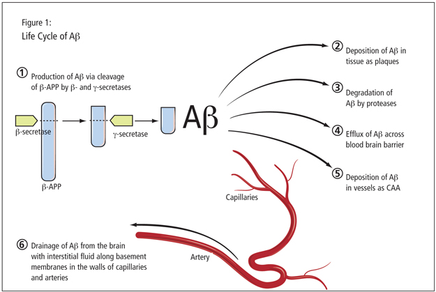
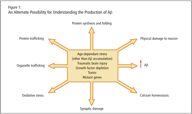
Dr. LeBlanc described age-dependent stresses, traumatic brain injury, growth factor depletion, toxins, and mutant genes as leading to increased Ab peptide. She alleged that the peptide may just be a consequence of cellular dysfunction such as protein trafficking, organelle trafficking, and oxidative stress (Figure 1). A critical experiment, she proposed, would involve altering g-secretase sites of the APP and studying the consequence. She stated that mutant genes would need to be expressed in absence of amyloid production to determine that cells are not affected in an amyloid-independent manner by the genetic mutation.

The fifth argument concerns whether Ab is a neurotoxic peptide; Dr. LeBlanc granted that this is amply demonstrated. Cytotoxicity may be associated with a very particular form of Ab, the oligomer. However, Dr. LeBlanc countered by noting that in research, cytotoxosis is induced at much higher levels than physiological concentrations. In addition, cell death is not widely evident in brains of patients with AD and is not correlated with the level of dementia experienced. Dr. LeBlanc claimed that intracellular Ab is a more compelling potential cause of AD.

The sixth argument addresses the finding that genetically engineered and transgenic animals carrying mutant AD genes generate more amyloid and develop AD pathology and cognitive impairment. These problems have been reversed by anti-amyloid immunotherapy. However, Dr. LeBlanc noted that an APP transgenic mouse with an additional mutation at the caspase C-terminal site was found to not have any cognitive impairment, despite retaining the ability to make the same amount of Ab peptide.

The final argument for the hypothesis, supported by research, is that amyloid perturbs the synaptic function. Synaptic proteins are downregulated in AD, and soluble Ab is able to dysregulate synapses. As cited by Dr. Lovestone, an Ab oligomer injected directly in the brains of mice causes LTP problems. However, Dr. LeBlanc asserted that studies are required to prove that amyloid overproduction is preceding the synaptic problems and not the other way around.

An Alternative to the Amyloid-Beta Hypothesis: Is Amyloid Production a Consequence of the Disease?
Dr. LeBlanc asked, What if amyloid is a consequence of the disease and that age-dependent stresses cause amyloid beta peptide and neuritic plaque accumulation, neurofibrillary tangles, neuritic degeneration, and synapse loss?

This has been the focus of the research conducted by Dr. LeBlanc and her colleagues over the last 14 years. Working with primary cultures of human neurons, they removed the growth factors by serum deprivation. The cultures make two to four times more Ab peptide, dependent on caspase-6. Caspase-6 is a protease that degrades other proteins when activated; it is normally present in the brain but not activated unless there is an insult. Dr. LeBlanc and colleagues designed an antibody that would recognize only the active form of caspase-6 in the brain of AD patients and found that it is associated with all neuropathological markers. To prove the enzyme would be active in AD brain, they made a second antibody directed against the tau protein cleaved by caspase-6, and found that the cleaved tau is present in pretangles, extra- and intracellular tangles, and neurophil threads. Some patients, she noted, have carpets of these threads full of tau cleaved by caspase-6. They went on to study a series of cases to investigate whether there was a correlation with cognitive impairment levels. Among their findings was that active caspase-6 was abundantly present in mild, moderate, and severe AD. They also found evidence that among noncognitively impaired subjects, there can be a large amount of neurophil threads stained for tau cleaved with caspase-6. This occurs in the absence of hyperphosphorylation of tau; Dr. LeBlanc stated that it seems to precede the other markers of tau hyperphosphorylation.

Dr. LeBlanc concluded that since there is evidence that the amyloid hypothesis does not fully account for AD pathology, research should address what could be done in addition to stopping the Ab peptide to prevent cognitive impairment.

Curing Alzheimer’s Disease: The Case of Tau and GSK-3

Curing Alzheimer’s Disease: The Case of Tau and GSK-3

Teaser: 

Click here to view the entire report from the 4th Canadian Colloquium on Dementia

Curing Alzheimer’s Disease: The Case of Tau and GSK-3

Speaker: Dr. Simon Lovestone, PhD, MRCPsych, Professor, Old Age Psychiatry, MRC Centre for Neurodegeneration Research; Departments of Old Age Psychiatry and Neuroscience, King's College London, Institute of Psychiatry, London, England, UK.

The second speaker, Dr. Simon Lovestone, deemed the title of his presentation “inflammatory.” Given the complexity of AD therapeutics, can one really talk about “curing Alzheimer’s”?
Most AD treatments, Dr. Lovestone noted, are symptomatic. Disease modification therapies directed at the pathogenesis (i.e., the amyloid therapies) may or may not be curative. Dr. Lovestone explicitly defined curative treatments as those that may restore function.

Dr. Lovestone observed that most think that people with AD have symptoms because they are losing neurons. He granted the veracity of this hypothesis and that neuronal loss indeed causes symptoms. However, this notion offers little hope for people with AD, for no treatment restores neurons, short of stem-cell therapy-a therapy still in its infancy. Given this fact, and the fact that there may be no available therapy for years to come that is able to do more than slow disease progression, it would seem that there is little cause for optimism on the part of the public or researchers.

However, Dr. Lovestone rejected pessimism and suggested that treatments to restore function are within grasp. Dr. Lovestone claims that there are areas in the amyloid cascade hypothesis where restoring function is a meaningful objective-that is, from a pathogenic viewpoint, there is room to ask, could dementia symptoms result from a failure of neuronal function rather than a loss of neurons?

Tau Aggregation and Hyperphosphorylation in AD

One potential area Dr. Lovestone identified concerns tau. Neurofibrillary tangles are made of protein and tau. Tau is aggregated and phosphorylated in AD, and in neurodegenerative conditions—the tauopathies—the expression of the protein itself is altered. Dr. Lovestone advocated shifting attention from the role of the synapses and focusing instead on the protein machinery in the cell body. These proteins need to be transported to the synapse and elsewhere via axonal transport. The neurofilaments and microtubules facilitate the axonal transport needed for neuronal function. Vesicles, containing transported proteins, are held together by microtubular-associated proteins. Tau is an axonal-specific microtubular protein needed for normal neural function.
Dr. Lovestone described a hypothesis that when tau is hyperphosphorylated, or when phosphorylation is increased, tau tends to be removed. Microtubules tend to collapse and tau, under certain conditions, aggregates when not bound to microtubules. Presumably, Dr. Lovestone stated, that aggregation results in the neurofibrillary tangles of AD.

GSK-3 and Neuronal Function

Following this, Dr. Lovestone had four points to investigate: one, whether glycogen synthase kinase-3 (GSK-3) might be a tau kinase; two, whether GSK-3 might be altered in AD; three, whether there is any evidence that tau phosphorylation results in altered neuronal function; and four, whether GSK-3 might regulate neuronal function (Figure 1).

Research suggests that there is a range of kinases, including GSK-3, that will phosphorylate tau. Tau phosphorylation is switched off when the neurons are treated with a GSK inhibitor, as demonstrated in a study that used lithium. GSK-3 inhibition regulates tau phosphorylation in neurons, as a 1999 study showed.1

Regarding the second point, as to whether GSK-3 is altered in AD, Dr. Lovestone noted that it is difficult to test this hypothesis without accessing the brains of AD patients. Work by Pei et al. and Hye et al. has demonstrated that GSK-3 is increased in AD brains and that activity of GSK-3 correlates with damage. Dr. Lovestone and colleagues have investigated peripheral circulating cells; research suggests that GSK-3 is increased in white matter cells in AD.

As to whether GSK-3 protein is itself the best evidence for alteration in AD, Dr. Lovestone answers in the negative, saying evidence will likely come from understanding GSK regulation. It is inhibited by Wnt/wingless signaling and insulin signaling. Interestingly, insulin resistance is a strong risk factor for AD.

Going into the genetic evidence, Dr. Lovestone noted that a series of studies relevant to GSK, focusing on mediators of insulin signaling, have emerged from diabetes research. A diabetes study replicated with people with AD found association with a number of genes on the signaling pathway. There is further evidence from whole genome studies: according to one 2007 study, Gab2 alleles modify AD risk in APOE4 carriers, Dr. Lovestone stated. He further cited several chromosome studies showing association of mediators of insulin signaling with AD. Such studies have offered tentative evidence that regulation of GSK-3 through Wnt and/or insulin might be genetically associated with AD.

Regarding evidence that altering tau phosphorylation might be associated with altered neuronal function, Dr. Lovestone mentioned relevant drosophila studies modeling tauopathies. In such models, the addition of either human tau protein or GSK-3 affected axonal transport through clumping of the vesicles-some clumped and moved, while others were stationary. This affected neuronal function in larvae and adult flies. The phenotype effect was a massive increase of tau when they overexpressed GSK-3. The treatment of larvae with a series of GSK-3 inhibitors (i.e., lithium) reversed the phenotype in the tau-only expressing drosophila. There is a GSK-3 dependent tau phenotype in the absence of tau aggregation. In reviewing their results, they looked closely for tau aggregation, and found no evidence of it.

As to his final query-whether manipulating GSK-3 alters neuronal function-Dr. Lovestone again turned to evidence from animal studies regarding long-term potentiation (LTP), an increase in synaptic strength that follows the stimulation of the chemical synapse. In these studies of memory potentiation, they took mice with memory deficits overexpressing GSK and induced LTP. In their studies, when GSK-3 was inhibited following LTP; the memory potentiation lasted for hours. Dr. Lovestone described GSK-3 as “a gate that needs to be shut for memory to be preserved.” This may serve as early indication of the potential for treatments that restore function; however, Dr. Lovestone acknowledged, GSK-3 may prove a difficult target for pharmaceutical therapeutics to hit.

Reference

  1. Lovestone S, Davis DR, Webster MT, et al. Lithium reduces tau phosphorylation: effects in living cells and in neurons at therapeutic concentrations. Biol Psychiatr 1999;45:995-1003.

Natural Human Antibodies for Alzheimer Disease

Natural Human Antibodies for Alzheimer Disease

Teaser: 

Click here to view the entire report from the 4th Canadian Colloquium on Dementia

The Alzheimer’s Disease Symposium: Immunotherapies, the Question of Cures, and the Amyloid Hypothesis

Moderator: John R. Wherrett, MD, FRCP(C), PhD, Professor Emeritus, Division of Neurology, University of Toronto; consultant in Neurology, Toronto Western Hospital and Toronto Rehabilitation Institute; member, Memory Clinic, Toronto Western Hospital, Toronto, ON.
A panel of four high-profile researchers discussed current horizons of research in Alzheimer’s disease and related dementias.

Natural Human Antibodies for Alzheimer’s Disease

Speaker: Dr. Norman Relkin, MD, PhD, Director, Cornell Memory Disorders Program, New York Presbyterian Hospital; Weill Cornell Medical College, New York.
Neurologist Dr. Norman Relkin detailed his and others’ work in the field of immunotherapy for Alzheimer’s disease (AD).

Noting that disease treatments tend to reflect prevailing pathological hypotheses, Dr. Relkin observed that current drug therapies for AD reflect the so-called cholinergic hypothesis of the previous few decades. Today, most studies focus on the role of beta-amyloid, and there are presently more than 30 trials underway that pursue the amyloid hypothesis. These research approaches encompass the transcription and translation of the APP gene, the production of amyloid monomer, the aggregation process, the formation of diffuse plaques in the brain, and the inflammatory processes that convert the formation of plaques in the brain associated with AD.

Dr. Relkin reminded listeners that immunotherapy is a class of treatments, not a single therapy. Immunotherapies include any approach that harnesses the actions of the humoral (B-cell mediated) and/or cellular (T-cell mediated) immune system. Immunotherapy initially came to the fore after Dr. Dale Schenk and colleagues found that an amyloid “vaccine” reduced the burden of plaques and memory loss in a transgenic mouse model of AD. The effect was demonstrated in the case of established plaques as well, despite the former thought that plaques were immutable.

The first use of antiamyloid immunotherapy in humans with AD showed similar clearing of amyloid plaques. However, due to a harmful T-cell mediated response, an unacceptably high number of patients developed meningoencephalitis. Subsequently, researchers sought to define and preserve the benefits of vaccination and eliminate the harmful response.

Dr. Relkin then described a developing approach called passive immunization, which involves supplying antibodies from an exogenous source, i.e., synthetic or animal. Dr. Relkin also drew attention to immunomodulation, a strategy that does not use the immune system directly but instead its chemical messengers, such as cytokines. All of the forms of immunotherapy he described are being pursued in current research.

Intravenous Immunoglobulin
Dr. Relkin further described a slightly different model of immunotherapy emergent over the last 4 years: intravenous immunoglobulin (IVIg), described in Figure 1.

Motivated by the safety concerns of amyloid immunotherapy, IVIg holds promise as something time-tested. IVIg is a pooled human Immunoglobulin G (IgG) preparation obtained by cold ethanol extraction from the plasma of thousands of healthy blood donors. It is already in use as a treatment for a number of immune deficiency disorders and other syndromes. IVIg contains natural antibodies, including antiamyloid antibodies.

Research data suggest that AD patients have fewer antiamyloid antibodies than nondemented individuals, making IVIg potentially valuable for such patients. IVIg has an established safety profile and is less likely than other immunotherapies to cause untoward events.

Dr. Relkin further detailed positive findings from his own research and that of Dr. Richard Dodel in Germany. Both studies’ Phase I data reflected a symptomatic benefit generated of IVIg over a 6-month period (these were add-on studies for patients under treatment with other agents), lowering the levels of amyloid in the blood. In the Relkin et al. study, a single IVIg low dose was followed by three higher doses. Each time the levels of antiamyloid antibodies were raised, there was a comparable impulse function in plasma amyloid curves. Dr. Relkin also presented data from his 18-month extended study. Significant results included marked decreases of the level of Ab40 and Ab42 in spinal fluid. Dr. Relkin then added a Mini-Mental State Exam measure to the study for safety purposes. He noted that the most robust response was seen among patients receiving the lowest doses of IVIg; a high number of low dose treatments also yielded positive results, in contrary to higher doses IVIg. Results were beyond expectations, the key finding being a disease stabilization effect over 18 months, Dr. Relkin stated. Given the positive interim results, they have announced they will be going to Phase III early in 2008.

Dr. Relkin noted some “paradoxes” in these data. Why do we have antibodies in our blood against Ab? Amyloid is a physiologic peptide we all produce from birth, for which the immune system should show tolerance. Researchers experimented with adding IVIg to Ab solutions in vitro: no binding response was exhibited. The response is mediated in the body by the effect of aggregation: amyloid can form soluble aggregates called oligomers, which are the targets of an antibody response. There is a peak of response corresponding to mid-sized oligomers, which means that the antibodies in IVIg are ignoring monomeric (benign) molecules and instead focusing on and binding to what Dr. Relkin described as very toxic species of reactive Ab. The “bottom line,” Dr. Relkin stated, is that the body is not indiscriminately fighting amyloid-it recognizes its toxic and misfolded forms.

The promise of this research, Dr. Relkin stated, is that oligomers are suspected to be the causative factors of a number of neurodegenerative diseases (AD, dementia in Down Syndrome, Lewy body dementia, Parkinson’s disease, Huntington’s, and others). These proteins circulate normally, but the oligomers represent pathological forms. This research suggests that there is an innate biological defense against neurodegenerative diseases, and IVIg holds promise as a tool to aid the fight (Table 1).

Alzheimer’s Disease: Treatments on the Horizon

Alzheimer’s Disease: Treatments on the Horizon

Teaser: 

Click here to view the entire report from the 4th Canadian Colloquium on Dementia

Alzheimer’s Disease: Treatments on the Horizon

Speaker: Dr. Serge Gauthier, MD, FRCPC, Director, Alzheimer Disease and Related Disorders Research Unit, McGill Centre for Studies in Aging, Montreal, QC.

Dr. Serge Gauthier focused his overview of current and future dementia treatments on the continuum of cognitive decline. He started with a “natural history of cognitive decline,” tracing cognitive deterioration’s trajectory from the earliest stages through profound impairment and death. The resulting picture is of a spectrum of cognitive abilities embracing the full range of health and pathology.

With this continuum in mind, Dr. Gauthier termed normal cognitive abilities as “pre-Mild Cognitive Impairment.” Such individuals might have subjective memory complaints, but these are undemonstrated in testing.

The next position on the spectrum was “Amnestic MCI” or “Predementia Alzheimer’s Disease.” This may be a prodromal phenomenon, Dr. Gauthier explained. These individuals he called “the forgetfuls,” noting that some but not all have pre-dementia Alzheimer’s disease (AD). Current symptomatic treatments for this stage include antidepressants and cholinesterase inhibitors (ChEI).
The choice of medication and the development of future treatments is connected to the concept of disease progression (Figure 1). He advised the audience to recall the difference between disease-modifying versus symptomatic drugs. With the latter, no improvement in the patient’s condition is expected, the criterion being only stabilization.

Dr. Gauthier cited a study by Winblad et al. from Neurology (2001) as evidence of donepezil’s benefit.1 He emphasized that this is a class effect: clinicians can expect improvement over baseline for 9-12 months for mild to moderate levels of impairment. Individuals with moderate to severe impairment living at home can expect improvement for about 6 months. In terms of cognition, patients will improve for anywhere from 6-12 months, depending on the severity of their cognitive decline.

Dr. Gauthier addressed the issue of combining agents, namely, memantine and donepezil in patients with mild dementia. He noted that studies have found additional improvement in cognition scores with the combined treatment over a period of 6 months.

In terms of agents on the horizon, Dr. Gauthier discussed the coming rivastigmine transdermal patch. The theoretical benefits, he stated, are clear. Recent studies confirm demonstrated benefit over baseline in MMSE scores. Similar benefits were demonstrated on the ADCS-ADL. The benefit with the patch is that higher doses may be achievable, without compromise to safety and tolerability.

Dr. Gauthier emphasized the value of Goal Attainment Scaling, as discussed by Dr. Kenneth Rockwood in the Canadian Medical Association Journal. Individual goal setting, in combination with galantamine, can be of value to patient care when real improvement in disease expression and progression is not expected, he noted.

The key idea, Dr. Gauthier stressed, is that symptomatic treatments for mild to severe stages of AD and related dementias are already available.

Modifying Disease among Mild AD Patients
Dr. Gauthier discussed disease modification among the patient group on the mild end of the spectrum of AD expression. In presenting details of two studies, he mentioned the caveat that the traditional study duration is sometimes inadequate to chart improvement among this treatment group. Eighteen months may be insufficient with mild AD patients.

Tramiprosate & Taranflurbil

Dr. Gauthier discussed treatment trials of the agent tramiprosate (Alzhemed®), an agent that binds to the soluble amyloid Ab. Phase III data from the clinical trial did not demonstrate statistical significance in favour of tramiprosate, with respect to the primary endpoints over 18 months of treatment. An advisory board of experts is currently reviewing data to see if benefit could have been measured differently. A European Phase III clinical trial is ongoing and could be modified based on North American findings.

The second agent Dr. Gauthier considered was taranflurbil (Flurizan®), a modulator of gamma secretase, currently in Phase II testing under lead investigator Dr. Sandra Black. The Phase II effort is to determine the most appropriate dosage. Patients in the study’s Canadian arm continued on the regime for an extra year. Key results showed that patients with mild AD who received a high dose of the medication obtained MMSE results indicating stabilization. Another surprise from the Phase II data was the statistically significant reduction of the psychiatric side effects. The protocol is ongoing for Phase III, and results are expected in December 2008.

Dr. Gauthier observed that there is room for improvement in the responder analysis: he offered that the 18-month baseline is not as useful a measure as it could be, and that a responder analysis would be useful. Further, delay to events-that is, the delayed emergence of significant psychiatric symptoms-would be a more meaningful measure of benefit, according to Dr. Gauthier. Disease-modifying drugs are under testing in mild AD, the primary hypothesis being that amyloid deposition is a major cause of the disease. The question remains, is this stage of dementia the best one for such pharmacotherapies?

A Closer Look at Mild Cognitive Impairment
Mild cognitive impairment (MCI), Dr. Gauthier emphasized, is neither a diagnosis nor a disease, but a way of defining cognitive deficit that has clinical utility. There will never be a drug for MCI, he stated. He presented results of a joint Canada-U.S. study comparing Vitamin E, donepezil, and placebo.2 The study found no difference in conversion rates at 3 years for any of the groups. At present, there is nothing to be given for MCI.

However, the data are different for predementia Alzheimer’s. Dr. Gauthier mentioned his recent position paper considering research criteria for the diagnosis of AD, published in 2007 in Lancet Neurology and advocating a change in the core diagnostic criteria.3

The ultimate effect of the changed criteria is to extend the determination of Alzheimer’s in a predementia stage. A diagnosis of the predementia stage of AD would become a clinical possibility and would be considered amenable to experimental treatments, with progression to dementia as a primary endpoint.

Conclusion: Controllable Risk Factors for Cognitive Decline
Dr. Gauthier predicted that control of risk factors will be integrated as part of the general stage-specific intervention schema. He noted specifically that education, dietary, and lifestyle factors have been correlated with cognitive effects. Incorporating measures to improve these factors will be integral to efforts to slow the conversion rate of pre-AD memory to dementia, and strategies to target them (such as interventions to control vascular risk factors) will be of key interest to researchers in coming years. This is fuelling study of agents such as ginkgo biloba. Dr. Gauthier advised predicted growing interest in the “triple approach” of diet (particularly the Mediterranean diet, shown to reduce risk by ~40%), exercise, and cognitive training. Overall, the focus of research and study for the next 5 years will likely reflect a preventive approach to cognitive impairment and the dementing syndromes.

References

  1. Winblad B, Engedahl K, Soininen H, et al. A 1-year, randomized, placebo-controlled study of donepezil in patients with mild to moderate AD. Neurology 2001;57:489-95.
  2. Peterson RC, Thomas RG, Grundman M, et al. Vitamin E and donepezil for the treatment of mild cognitive impairment. N Engl J Med 2005;352:2379-88.
  3. Dubois M, Feldman HH, Jacova C, et al. Research criteria for the diagnosis of Alzheimer’s disease: revising the NINCDS-ADRDA criteria. Lancet Neurol 2007;6:734-46.

Working Effectively with Community Partners in Dementia Care

Working Effectively with Community Partners in Dementia Care

Teaser: 

Click here to view the entire report from the 4th Canadian Colloquium on Dementia

Working Effectively with Community Partners in Dementia Care

Speaker: Dr. Carole A. Cohen, MD, FRCPC, Associate Professor, Department of Psychiatry, University of Toronto; Clinical Director, Community Psychiatric Services for the Elderly, Sunnybrook and Women’s College Health Sciences Centre; Toronto, ON.

Dr. Carole Cohen, a geriatric psychiatrist and academic, explored the wider “systems issues” associated with providing care to individuals diagnosed with dementia. These individuals’ care needs cannot be met solely within primary care. The key question, according to Dr. Cohen, is who are the community partners that one should approach?

Dr. Cohen described a pivotal shift in the conceptualization of dementia care that accompanies viewing the condition as a chronic disease. When dementia is considered a chronic condition (as outlined in Table 1), one may think more laterally and flexibly about the diversity of potential care needs, and about creating key partnerships-be they clinician/clinician partnerships or clinician/community partnerships. Such alliances can compensate for the challenges encountered in managing dementia in primary care.

Patient Needs
Patient needs are likely to include information on their diagnosis and appropriate course of treatment, educational information, supportive resources, psychological care, and decisional support as impairment increases. Inherent to the challenge of providing for these needs is that they will vary among a diverse patient population. Needs should also accord with dementia type and be tailored to each syndrome.

Many clinics are not equipped to address the full range of patient needs, and that, according to Dr. Cohen, is where community alliances help fill the breach.

Caregiver Needs
“What’s good for caregivers is good for patients,” asserted Dr. Cohen. If clinicians can help caregivers to mobilize social support, formal and informal, patient needs will also be met. The objective in meeting caregiver needs-which include education and information, social support, and assistance in mobilizing that support-is to fortify the wider circle of care around the individual with dementia.

Dr. Cohen noted that entities such as day programs, respite programs, and support groups are underused. Not only may caregivers be unaware of them, but many people do not think of themselves as caregivers and therefore do not seek out such support. They may see themselves simply as a son or daughter aiding an ailing parent. Such individuals, however, are at high risk for burnout. As Dr. Cohen described, what helps caregivers to avoid depression and burnout, will allow them to cope longer and better assist the individual with dementia. Research has shown the effectiveness of individualized plans, plans that have multiple strategies, training, information, and ongoing relationships-for the dementing illnesses may last 15 or 20 years.

Putting Dementia Care in Context
Given the foregoing, Dr. Cohen questioned how traditional care models will help the burgeoning dementia population. Dr. Cohen asked the audience to consider the broad context of dementia care. She urged listeners to “think beyond hospital walls,” to be cognizant of the “vast literature” on chronic care across many illnesses/conditions, and to recognize that many tenets for other models (diabetes, for example) will work for dementia.

An interesting model for dementia from chronic disease management is termed the “prepared practice team,” a multidisciplinary team aimed at providing broad psychosocial interventions. Supplemental strategies such as the creation of e-records, interchange of information, and coordination of team care result in an informed and empowered patient and family.

Borrowing from the Chronic Care Model

Two key parts of the chronic care model Dr. Cohen encouraged listeners to consider were facilitating self-management support and community resources (Figure 1).

Self-management could better support patients with dementia as well as their families. Self-care complements the care provided by the professional team and facilitates training to cope with the debilitating aspects of dementia, as well as promotes the use of decisional aids. Actively involving patients in their care is empowering when facing a disease bound to erode their problem-solving and decision-making capacities. Patients should be assisted in locating and using available resources and partnerships.

Disease self-management empowers individuals to become more actionable. Self-management mobilizes support, and this idea has led to the creation of programs such as cognitive memory rehabilitation services. The key, Dr. Cohen stated, is finding community resource partners to promote this kind of action. She advocates the use of case managers and family health teams. Clinicians need to ask what they can do to focus on and promote those community partnerships. Dr. Cohen used the example of an FTD daycare program at Baycrest Centre in north Toronto; she lamented that many dementia care programs are developed but underused. Patients and their caregivers need clinician encouragement to avail themselves of community programs.

Dr. Cohen suggested that clinicians might partner with caregiver resource centers, Alzheimer’s groups, and similar organizations.

Practical Suggestions for Clinicians

Dr. Cohen offered concrete suggestions for clinicians working with patients with dementia. She again advocated adopting the chronic care model for dementia. She urged them to work to better identify patient-caregiver needs. Identifying community partnerships, such as with “Meals on Wheels,” will facilitate patient care. She also urged clinicians to integrate case managers to serve as the “glue” connecting the levels of support and care.

Dr. Cohen concluded with Canadian examples of the kind of chronic care principles and community partnering she advocated. She described the FirstLink program, which is being implemented across Canada. This program provides an individual with a new dementia diagnosis a direct referral to a FirstLink clinic, which in turn gives the Alzheimer’s Society permission to call and offer available programs, services, and training. She described the founding of this program as a boon to busy MDs.

Regional networks and programs, such as the coordination of care between the Alzheimer’s Society and geriatricians in Prince Edward Island, as well as the networks of the British Columbia-based dementia clinics and community partnerships, stood out for Dr. Cohen as the kind of interventions beyond clinic walls that serves patient needs. She encouraged listeners to continue to think in these new directions, as the extent and gravity of the dementing diseases will demand the utmost of the health care system.

The Latest Research in Dementia Care: Views from the 4th Annual Canadian Colloquium on Dementia

The Latest Research in Dementia Care: Views from the 4th Annual Canadian Colloquium on Dementia

Teaser: 

Click here to view the entire report from the 4th Canadian Colloquium on Dementia

The Latest Research in Dementia Care: Views from the 4th Annual Canadian Colloquium on Dementia

The 4th Annual Canadian Colloquium on Dementia: An Overview

Speaker: Dr. Ron Keren, MD, FRCPC, 4th CCD Chair; Clinical Director, University Health Network and Whitby Mental Health Centre Memory Clinics; Assistant Professor, University of Toronto, Toronto, ON.

At present, an estimated 450,000 Canadians over 65 have Alzheimer’s disease (AD) or other related dementias. The incidence and prevalence of AD and related dementias is on the rise as the population continues to age. The expected scope and impact of the condition, and the desire to share research findings on the diagnosis and management of dementia, has led to the convening of an annual colloquium for Canadian and international dementia experts known as the Canadian Colloquium on Dementia.

The 4th Annual Canadian Colloquium on Dementia, held October 18-20, 2007 in Vancouver BC, brought together a large, expert faculty of Canadian and international speakers. The conference attracted a record number of residents and fellows compared with years past. As noted by Dr. Ron Keren, who chaired the colloquium in cooperation with co-chair Dr. Howard Feldman, the annual gathering provides a setting for exploring both clinical applications of latest research in dementia care as well as a consideration of future trends in the diagnosis and treatment of a burgeoning condition. Given both the increased attendance and record number of abstracts received by the colloquium’s organizers for the 2007 meeting, Dr. Keren described this interest in the subject as “boding well for the future of dementia care” in his opening remarks.

This report offers a closer look at select presentations from the colloquium that addressed key issues related to the diagnosis and management of dementia.

Mild Cognitive Impairment: What Is It and Where Does It Lead?

Mild Cognitive Impairment: What Is It and Where Does It Lead?

Teaser: 


Lesley J. Ritchie, MSc, Department of Psychology, Centre on Aging, University of Victoria, Victoria, BC.
Holly Tuokko, PhD, Department of Psychology, Centre on Aging, University of Victoria, Victoria, BC.

Mild cognitive impairment (MCI) is an intermediary stage in the cognitive continuum from normal aging to dementia. Six to 48% of individuals with MCI are estimated to develop dementia.1 As such, the conceptualization and operationalization of MCI present unique opportunities for the development and implementation of strategies to prevent or delay the conversion to dementia. Despite the lack of a “gold standard” case definition for MCI, information gathered from neuropsychological assessment may inform a diagnosis of MCI based on clinical judgment, as impaired performance on several neuropsychological measures is predictive of conversion to dementia for persons exhibiting cognitive decline but who are not demented.
Key words: mild cognitive impairment, dementia, conversion, neuropsychology, predictors of dementia.

The Role of the Neurologic Examination in the Diagnosis and Categorization of Dementia

The Role of the Neurologic Examination in the Diagnosis and Categorization of Dementia

Teaser: 

John R. Wherrett, MD, FRCP(C), PhD, Professor Emeritus, Division of Neurology, University of Toronto; consultant in Neurology, Toronto Western Hospital and Toronto Rehabilitation Institute; member, Memory Clinic, Toronto Western Hospital, Toronto, ON.

Nonneurologist practitioners faced with the diagnosis of dementia cannot be expected to conduct the detailed assessments for which neurologists are trained. Nonetheless, they should be able to diagnose the most common forms of neurodegenerative dementia and identify individuals that require more detailed neurologic workup. A neurologic examination algorithm is described that allows the practitioner, in a stepwise and efficient manner, to elicit findings that distinguish the main categories of neurodegenerative and vascular dementia, namely, Alzheimer’s disease, dementia with Lewy bodies, vascular dementia, and frontotemporal lobar degenerations. Patients are assessed for gait, frontal signs, signs of parkinsonism, signs of focal or lateralized lesions, neuro-ophthalmologic signs, and signs characteristic of frontotemporal lobar degeneration.
Key words: neurologic, examination, neurodegenerative, dementia, diagnosis, gait, frontal dysfunction, cognitive impairment.

Assessing Patients Complaining of Memory Impairment

Assessing Patients Complaining of Memory Impairment

Teaser: 


Mario Masellis, MSc, MD, FRCPC, Clinical Associate & Research Fellow, L.C. Campbell Cognitive Neurology Research Unit, Division of Neurology, Department of Medicine, Sunnybrook Health Sciences Centre, University of Toronto, Toronto, ON.
Sandra E. Black, MD, FRCPC, Brill Professor of Neurology, L.C. Campbell Cognitive Neurology Research Unit, Division of Neurology, Department of Medicine, Sunnybrook Health Sciences Centre, University of Toronto, Toronto, ON.

Cognitive impairment occurs along a continuum from mild subjective memory complaints occurring during the normal aging process to severe memory and other cognitive deficits due to dementia, the most common subtype being mixed Alzheimer’s disease and vascular dementia. Due to the significant growth of the older adult population, the incidence of dementia is on the rise and is posing significant challenges for health care systems worldwide. Primary care practitioners are on the front lines of this battle against dementia and will play an increasingly important role in the early identification of disease. Cognitive screening tests are helpful in detecting people in the early stages of dementia and facilitate further clinical and diagnostic evaluations. Primary care practitioners should aggressively treat known cardiovascular risk factors for dementia and institute early symptomatic therapy when appropriate.
Key words: dementia, cognitive screening test, cognitive reserve, neuroimaging, biomarkers.