Advertisement

Advertisement

aging

Neurobiologie du vieillissement

Neurobiologie du vieillissement

Teaser: 


Neurobiologie du vieillissement

Conférencier : David F. Tang-Wai, MDCM, FRCPC, Professeur adjoint (en neurologie), Université de Toronto ; University Health Network Memory Clinic, Toronto, ON.

Le Dr David Tang-Wai a passé en revue les mécanismes de base du vieillissement humain, et a décrit en détail l’application des théories du vieillissement aux troubles neurodégénératifs, en particulier la maladie d’Alzheimer (MA). Il a examiné la neuropathologie de la MA et ses facteurs étiologiques.

Le Dr Tang-Wai, qui a salué l’intervention des conférenciers précédents, a fait remarquer que le vieillissement repose sur des mécanismes multiples, plutôt que simples, et que tous ces facteurs impliqués dans le vieillissement neurologique et ses désordres – depuis la génétique jusqu’au stress oxydatif, le dysfonctionnement mitochondrial, et l’accumulation des cellules sénescentes – sont des processus interagissant et inextricables.

Énumérant certains des principaux changements du système nerveux avec l’âge, le Dr Tang-Wai a mentionné l’importance des changements physiologiques liés au vieillissement, comme l’atrophie cérébrale, la diminution des synthèses catécholaminergique et dopaminergique, la baisse des réflexes de redressement, et la réduction du sommeil de stade 4. Ceci se manifeste sur le plan clinique par une démarche altérée/ralentie, un balancement du corps et une certaine étourderie, ainsi que par des troubles du sommeil (p. ex., de l’insomnie). Cependant, ces changements doivent être distingués des états suggestifs de processus pathologiques tels que la démence, la dépression, les troubles du mouvement comme la maladie de Parkinson, les chutes et l’apnée du sommeil, dont aucun n’est un attribut normal du processus de vieillissement.

Des études récentes confirment qu’il existe un certain déclin cognitif universel accompagnant le vieillissement, déclin qui débute au début de l’âge adulte. L’efficacité et l’aptitude mentales atteignent leur pic vers la fin de la vingtaine, puis entament un déclin.
Puis le Dr Tang-Wai a fait un tour d’horizon de la MA, en notant que l’âge est le facteur de risque le plus important de la maladie, ce risque doublant après 65 ans. La maladie d’Alzheimer est la forme la plus courante de la démence de l’adulte, mais elle risque d’être bientôt dépassée par la démence mixte Alzheimer-vasculaire. Sur le plan pathologique, le cerveau subit dans la MA une atrophie globale, un élargissement des sillons corticaux, une réduction des gyri et un élargissement de la scissure de Sylvius (Figure 1).


L’évolution de la MA à l’échelle microscopique aboutit à une perte synaptique et neuronale. Finalement apparaissent les signatures pathologiques de la MA, en particulier les corps d’Hirano, les enchevêtrements neurofibrillaires, la dégénérescence granulovacuolaire et les plaques séniles.

Le Dr Tang-Wai a récapitulé la formation des enchevêtrements, qui sont le résultat de l’hyperphosphorylation des protéines tau (la protéine tau présente 6 isoformes qui s’apparient et s’agglomèrent, formant finalement des enchevêtrements), ainsi que des plaques séniles (dépôts extracellulaires d’amyloïde). Il a présenté en détail les multiples types de plaques séniles, dont la plaque diffuse du vieillissement, qui peut être observée chez la personne âgée non démente ; la plaque neuritique avec un noyau amyloïde dense ; et, avec le temps, une plaque « dégénérée » qui laisse un important noyau amyloïde. Ce processus débute dans l’hippocampe ; c’est donc la mémoire qui est touchée la première.

Discutant de la distribution des plaques et des enchevêtrements, ainsi que des stades de Braak, le Dr Tang-Wai a décrit les dérèglements initiaux de la mémoire, passant par le cortex associatif et affectant les systèmes sensoriels et moteurs au cours des derniers stades. La mention de l’hypothèse de l’amyloïde bêta (Ab) a mené à une discussion des principaux facteurs de risque de MA, notamment l’allèle de l’apolipoprotéine E (qui accélère le déclin cognitif chez la personne âgée cognitivement normale et qui est associé à une diminution de la vitesse de traitement et de l’apprentissage, ainsi qu’à une réduction du métabolisme du glucose), l’hypertension, l’obésité, l’hypercholestérolémie et le diabète. Le Dr Tang-Wai a porté une attention particulière au diabète en tant que facteur de risque dont on mesure à nouveau de plus en plus l’importance. Il a fait remarquer que l’insuline influe sur la mémoire par sa modulation de la structure et de la fonction synaptique, sa potentialisation à long terme et ses modifications des taux de neurotransmetteurs du SNC. L’insuline a des effets protecteurs sur le cerveau. Les états pathologiques d’insulinorésistance comprennent l’hyperinsulinémie, qui réduit le transport d’insuline à travers la barrière hémato-encéphalique et diminue les concentrations et l’activité de l’insuline dans le cerveau. La réduction du signal de l’insuline dans le cerveau est associée à une augmentation de la phosphorylation des protéines tau et à des concentrations plus élevées d’Ab, qui sont les signes pathologiques directs de la MA.

L’obésité et l’hypercholestérolémie ont comme effets indésirables d’augmenter l’insulinorésistance ainsi que d’augmenter les acides gras libres (AGL). De très fortes oscillations de taux d’AGL élevés entraînent une cascade d’effets indésirables. Elles inhibent les enzymes de dégradation de l’insuline, diminuant ainsi la clairance de l’Ab, et stimulant l’assemblage de la plaque amyloïde et des filaments des protéines tau. Les acides gras libres peuvent également induire une inflammation, qui stimule indirectement la formation de la plaque amyloïde et les enchevêtrements neurofibrillaires.

Parmi les principaux facteurs de risque de MA, on compte également l’hypertension de la cinquantaine, qui implique aussi une résistance à l’insuline et le risque d’un dépôt important d’Ab ; et le stress oxydatif, auquel le cerveau est particulièrement sensible, car il est largement composé de lipides facilement oxydables, il a un taux élevé de consommation d’oxygène (un quart de l’oxygène consommé va directement au cerveau), et il est dépourvu de défenses antioxydantes fortes, a encore fait remarquer le Dr Tang-Wai. Le lien entre la MA et le stress oxydatif est surtout associé à l’augmentation de l’oxydation dans le cerveau avec le vieillissement.
La dysfonction mitochondriale, ou dysfonction énergétique, joue également un rôle dans la MA. Les recherches ont montré que l’Ab peut interagir avec les mitochondries et induire une dysfonction mitochondriale. Celle-ci accélère les processus indésirables associés à la neurodégénérescence (altération de l’homéostasie calcique, génération de RLO, glycol-oxydation pouvant accélérer l’agrégation des Ab et accroître la prolifération de microglie ; mutation de l’ADN ; et altérations du traitement des protéines tau et Ab). De plus, les changements oxydatifs dans la MA sont associés aux enchevêtrements neurofibrillaires (et la théorie veut que les effets des dommages oxydatifs perturbent le neurone lui-même), ce que démontre la présence de carbonyles protéiques, de produits de la peroxydation des lipides, et des produits ultimes d’une glycation avancée.

On met de plus en plus l’accent sur le rôle de l’inflammation dans la MA par l’intermédiaire des effets synergiques de trois éléments principaux : la microglie, les astrocytes et les neurones. Les éléments inflammatoires relatifs à la neuroinflammation dans la MA comprennent la microglie et les astrocytes, qui tous deux génèrent indirectement des Ab. La microglie entoure le neurone et peut produire des radicaux libres de l’oxygène et du monoxyde d’azote, entraînant une neurodégénérescence. Les astrocytes peuvent induire une neurodégénérescence par la production de ra-dicaux libres de l’oxygène, la dégradation des Ab, et la production de cytokines et de chimiokines, favorisant encore plus l’implication de la microglie. Les neurones endommagés peuvent eux-mêmes produire des cytokines et des chimiokines, et augmenter les protéines CRP et amyloïdes P, qui activent alors le système du complément. Finalement, l’Ab peut elle-même être un facteur incitatif de la genèse des astrocytes et de l’activation microgliale. En conséquence de l’augmentation de la production de radicaux libres de l’oxygène et de la production de cytokines et de chimiokines, il y a phagocytose et dégradation de l’Ab. La protéine tau peut avoir une influence sur l’activation des systèmes du complément. En cas de régulation positive de la microglie, il peut y avoir une baisse de la dégradation de l’Ab et une baisse de l’insuline, induisant une intolérance au glucose, ce qui peut entraîner une sécrétion d’Ab. L’effet global produit boucles sur boucles de forces couplées.

Pour produire le résultat final de la pathologie de la MA, les principaux facteurs de risque et les facteurs génétiques doivent être en place. L’inflammation et le dommage oxydatif sont multisystémiques. Les processus relatifs à la pathologie de la MA présentés ici, a précisé le Dr Tang-Wai, s’appliquent à tous les processus neurodégénératifs ; ces conformations et agrégations de protéines ainsi que l’inflammation sont présentes dans les maladies de Parkinson et de Creutzfeldt-Jakob, ainsi que dans la sclérose latérale amyotrophique familiale.

Opening Ceremonies:Welcome and Presidential Address

Opening Ceremonies:Welcome and Presidential Address

Teaser: 


Opening Ceremonies: Welcome and Presidential Address

Speakers: Andrew D. Baker, MD, FRCPC, Consultant, Internal Medicine and Geriatrics, Toronto Rehabilitation Institute, Queen Elizabeth II Campus; Consultant, Geriatrics, Toronto Rehabilitation Institute, University Campus; Active Staff, Internal Medicine and Geriatrics. St. Joseph’s Health Centre, Chair, Section of Geriatric and Long Term Care, O.M.A Toronto, ON; Jennifer Ingram, MD, FRCPC, Founder, Kawartha Regional Memory Clinic, Peterborough, ON; Consulting Geriatrician, Whitby Mental Health Centre Memory Clinic, Whitby, ON; Chris Frank, MD, FCFP, Associate Professor, Department of Medicine, Queen's University; Clinical Director, Specialized Geriatrics, Providence Care, Kingston, ON.

The 29th Canadian Geriatrics Society (CGS) Annual Scientific Meeting launched with welcoming remarks delivered by its co-chairs, Dr. Andrew Baker and Dr. Jennifer Ingram. Dr. Baker is in full-time geriatrics and internal medicine practice at St. Joseph’s Health Centre in Toronto. Dr. Baker is the current OMA head of the section for Geriatrics and Long-Term Care and has been chief of geriatrics at St. Joseph’s since 2005. Dr. Ingram is a specialist in internal medicine and geriatric medicine currently practicing in Peterborough, Ontario. Dr. Ingram founded the Kawartha Regional Memory Clinic, a consultative service. Dr. Ingram also serves as consulting geriatrician at the Whitby Mental Health Centre’s Memory Clinic.

Dr. Baker introduced Dr. Chris Frank, outgoing President of CGS, who offered his own welcome to attendees. Dr. Frank, associate professor in the Department of Medicine at Queen’s University and director of the Care of the Elderly residency program, as well as Clinical Director of Providence Care Specialized Geriatric Services, reflected on the three themes that encompassed the efforts of CGS members during his presidency: the need for collaborative work with other organizations devoted to seniors’ health and well-being; the necessity to mobilize the intelligence and energy of the Society’s able members given the Society’s small size; and the commitment to excellence in patient care.

All three aims were evident in reviewing the previous year’s accomplishments, he noted: the Society has worked collaboratively with the Canadian Association of Retired Persons, the Canadian Pensioners Concerned, the Canadian Academy of Geriatric Psychiatry, the Health Care of the Elderly Committee of the College of Family Physicians, and the National Initiative for Care of the Elderly, among other organizations, to develop the Geriatric Education and Recruitment Initiative (GERI). The collaborative’s mandate is to improve the image of caring for seniors and to attract people to the field. Dr. Frank stated that GERI has a funded project to develop geriatric interest groups at health science centres and has worked with the media and related businesses to improve the representation of aging. Ongoing activities include efforts to secure grant monies to fund geriatric education in Africa.

As for CGS members’ commitment to senior care, he noted the multitude of voices that rose in response to the journal Geriatrics & Aging’s “Call to Arms,” which sought opinions on the value of geriatrics. Most respondents equated geriatrics with excellence in medical care.

Dr. Frank emphasized the Society’s pleasure at bringing multiple health care organizations together for the meeting to work collaboratively to promote excellence in eldercare. He encouraged attendees to incorporate the information and initiatives discussed into their professional interactions, and to build and maintain professional linkages. He emphasized his optimism about the growing attraction of geriatrics, noting the CGS membership was at an all-time high. Increasingly, core competencies in geriatric care are successfully being introduced into medical training, as those who map curricula are seeing the need to prepare practitioners for burgeoning numbers of aging adults. Dr. Frank expressed hope that the meeting would be of great utility in helping professionals to care for seniors in their locale.

Keynote Address

Keynote Address

Teaser: 

Keynote Address

Speaker: Her Worship Hazel McCallion, Mayor of Mississauga

Dr. Jenny Ingram introduced Her Worship Hazel McCallion, Mayor of Mississauga. Calling her “a true pioneer,” Dr. Ingram stated that after 11 consecutive elections and 31 years in office, Mayor McCallion is the longest-serving elected municipal politician in Canada, an inspiration to women executives, and has achieved a success that community leaders everywhere wish to parallel. Given her longevity and vivacity, Dr. Ingram observed, Mayor McCallion personifies the Canadian Geriatrics Society’s motto “Adding years to life and life to years.”

Mayor McCallion acknowledged that the challenges encountered by seniors are great, quoting Bette Davis: “Old age is no place for sissies,” and called CGS “a great organization” that has served older people well. However, the mayor also adheres to the adage, “You’re only as old as you think” and noted that while she is 88 years old, she experiences the same overall good health and vigour she felt in her thirties. She acknowledged that genetics might be thanked, but is of the conviction that sound living and preventive measures achieved by wise health habits is where most credit is due.

Mayor McCallion offered Mississauga as an example of a city that both recognizes the challenges that can accompany aging and accommodates them. She strives to promote seniors’ unique contribution to civic and social life. The city commissioned a study to devise ways to prepare for an expected shift in demographics that mirrors changes in other Canadian municipalities and many other communities worldwide. Over the next 20 years, the portion of city residents aged 55 and over is expected to rise to close to 40%, and a strategic plan to respond to this challenge, a 10-year planning study called the Older Adult Project, has been undertaken.1

Mississauga is investigating elder-friendly modifications to the community, which aim to facilitate active and healthy living. The study is examining improving accessibility, public transit and other mobility choices, concentration of vital services, public spaces and parks, and availability of community and recreation programs, among other areas. The overall principle is to ensure that “services, facilities, and programs are more accessible and responsive to the specific needs of older persons.” Additional focus is on features that enable seniors to remain physically active; in 2008, Mayor McCallion bicycled seven kilometres to Mississauga City Hall to raise awareness about the importance of remaining fit.

The mayor concluded her address by sharing poems and anecdotes from which she has drawn inspiration and support. Her final offer of advice concerned the importance of a positive attitude to how one experiences advanced age. However, she made a special point of acknowledging the essential services that geriatricians provide to seniors. She thanked the Canadian Geriatrics Society for its 29 years of service and its efforts to “put older citizens back into action.”

  1. Online at http://www.mississauga.ca/ file/COM/Old_Adult_Report_1.pdf

Neurobiology of Aging

Neurobiology of Aging

Teaser: 


Neurobiology of Aging

Speaker: David F. Tang-Wai, MDCM, FRCPC, Assistant Professor (Neurology), University of Toronto; University Health Network Memory Clinic, Toronto, ON.

Dr. David Tang-Wai presented a review of the basic mechanisms of human aging and offered a detailed application of the theories of aging to neurodegenerative disorders, foremost Alzheimer’s disease (AD). He discussed the neuropathology of AD and its causative factors.

Dr. Tang-Wai, acknowledging the previous speakers, noted that aging comprises multiple rather than single mechanisms, and all of those factors concerned in neurological aging and its disorders—from genetics to oxidative stress to mitochondrial dysfunction and accumulation of senescent cells—are interacting and inextricable processes.

Enumerating some of the key changes to the nervous system with age, Dr. Tang-Wai mentioned the import of age-related physiological change, such as brain atrophy, decreased catecholaminergic and dopaminergic synthesis, decreased “righting” reflexes, and decreased stage 4 sleep. Clinically these manifest in altered/slowed gait, body sway and forgetfulness, along with sleep disturbances (e.g., insomnia). However, these changes should be distinguished from conditions suggestive of disease processes, such as dementia, depression, movement disorders such as Parkinson’s, falls, and sleep apnea, none of which are normal accompaniments to the aging process.

Recent studies confirm that some cognitive decline is universal with advancing age, a decline that begins in early adulthood. Mental efficiency and capacity peak by the late 20s and then decline.

Moving on to an overview of AD, Dr. Tang-Wai noted that age is the biggest risk factor for the disease, the risk for which doubles beyond age 65. Alzheimer’s is the most common form of adult-onset dementia, but he noted that it may soon be surpassed by mixed Alzheimer-vascular dementia. What happens to the brain pathologically in AD is global shrinkage, widened sulcal margins, narrowed gyri, and increased sylvian fissure (Figure 1). As AD progresses at the microscopic level, synaptic and neuronal loss follow. Finally, pathological hallmarks of AD appear, including Hirano bodies, neurofibrillary tangles, granulovacuolar degeneration, and senile plaques.


He briefly reviewed the formation of tangles, which are the result of hyperphosphorylated tau (tau has 6 isoforms that pair up and conglomerate, ultimately forming tangles), as well as senile plaques (extracellular deposits of amyloid). He detailed multiple types of senile plaque, including diffuse plaque of aging, which may be seen in the nondemented older adult; neuritic plaque with a dense amylytic core; and with time “burned out” plaque that leaves a prominent amyloid core. This process begins in the hippocampus; thus, memory is first affected.

Discussing distribution of plaques and tangles as well as Braak staging, Dr. Tang-Wai described AD’s initial impairments in memory, progressing through the association cortex, and affecting sensory and motor systems in late stages. A mention of the amyloid beta (Ab) hypothesis prompted review of the predominant risk factors for AD, including Apolipoprotein E allele (accelerates cognitive decline in cognitively normal older adults and is associated with decreased processing speed and learning as well as reduced glucose metabolism), hypertension, obesity, hypercholesterolemia, and diabetes. Dr. Tang-Wai gave particular consideration to diabetes as a risk factor whose importance is under new and increased scrutiny. He noted that insulin influences memory by modulation of synaptic structure and function, long-term potentiation, and alterations of CNS levels of neurotransmitters. Insulin’s effects on the brain are protective. Insulin-resistant states include hyperinsulinemia, which reduces insulin transport across the blood-brain barrier and lowers insulin levels and activity in the brain. Reduced brain insulin signaling is associated with increased tau phosphorylation and higher Ab levels, which are the direct pathological signs of AD.

Obesity and hypercholesterolemia involve the adverse effects of increased insulin resistance as well as increased free fatty acids (FFA). Elevated FFA involve cascading adverse effects. They inhibit insulin-degrading enzymes, thus decreasing Ab clearance, stimulating assembly of amyloid plaque and tau filaments. Free fatty acids may also induce inflammation, which indirectly stimulates amyloid formation and neurofibrillary tangles.

Other key risk factors for AD include midlife hypertension, also involving insulin resistance and the risk of elevated Ab deposition; and oxidative stress, to which the brain is distinctly vulnerable as it is largely composed of easily oxidized lipids, has a high oxygen consumption rate (one-quarter of the oxygen consumed goes directly to the brain), and lacks strong antioxidant defenses, Dr. Tang-Wai noted. The link between AD and oxidative stress relates primarily to the increase in oxidation in the brain with aging.

Mitochondrial dysfunction, or energy dysfunction, is also implicated in AD. Research shows that Ab can interact with mitochondria and cause mitochondrial dysfunction. Mitochondrial dysfunction accelerates adverse processes associated with neurodegeneration (altered calcium homeostasis, ROS generation, glycol-oxidation, which can accelerate Ab aggregation and enhance proliferation of microglia; DNA mutation; and alterations of tau and Ab processing). Further, the oxidative changes in AD are associated with neurofibrillary tangles (and it has been theorized that effects of oxidative damage affect the neuron itself) as supported by the evidence of the presence of protein carbonyls, products of lipid peroxidation, and advanced glycation end products.

Focus is increasingly trained on the role of inflammation in AD through the synergistic effects among three major elements: microglia, astrocytes, and neurons. Inflammatory components related to AD neuroinflammation include microglia and astrocytes, both of which indirectly generate Ab. Microglia surround the neuron, and can produce reactive oxygen species and nitric oxide, leading to neurodegeneration. Astrocytes can produce neurodegeneration through production of reactive oxygen species, degradation of Ab, and production of cytokines and chemokines leading to more involvement of microglia. Damaged neurons themselves can also produce cytokines and chemokines, and increase CRP and amyloid P, which then activate the complement system. Finally, Ab itself can be an inciting factor for the genesis of astrocyte and microglial activation. Because there is increased production of reactive oxygen species and production of cytokines and chemokines, the result is phagocytosis and degradation of Ab. The tau protein can influence activation of complement systems. Microglia if upregulated can lead to decreases in Ab degradation and decreased insulin, leading to glucose intolerance, which can lead to Ab secretion. The sum effect is loops upon loops of compounding forces.

In order to produce the end result of AD pathology, key risk factors and genetic factors must be in place. Inflammation and oxidative injury are multisystemic. The processes he detailed in connection with AD pathology, Dr. Tang-Wai observed, apply to all neurodegenerative processes; these protein conformations/aggregations and inflammation are present in Parkinson’s disease and Creutzfeldt Jakob, as well as familial amyotrophic lateral sclerosis.

Normal Aging Part 1: Cardiovascular, Respiratory, Gastrointestinal

Normal Aging Part 1: Cardiovascular, Respiratory, Gastrointestinal

Teaser: 


Normal Aging Part 1: Cardiovascular, Respiratory, Gastrointestinal

Speaker: Karen Fruetel, M.Ed, MD, FRCPC, Division of Geriatric Medicine, University of Calgary, Calgary, AB.

Dr. Karen Fruetel emphasized in her review of the physiological changes accompanying aging that attributes of “normal aging” have been derived from clinical studies. The studies are either cross-sectional, meaning that health markers are derived from comparisons between predefined aging cohorts, or are longitudinal studies that follow the same individuals over a number of years. Both types have their biases. Cross-sectional studies may falsely overlook confounders (e.g., comparing 20-year-old and 80-year-old study participants may overlook the fact that the latter were more likely to have smoked than today’s youth). Data from longitudinal studies are generally thought to be more robust, but such data may reveal selection biases (e.g., higher educational and/or socioeconomic status). Obtaining information on the true physiological consequences of aging is a challenge.

Dr. Fruetel commenced with a review of aging’s effects on the cardiovascular system. Autopsy studies suggest that advanced age is associated with increased left ventricular (LV) mass. Studies also show increased myocyte size and decreased number, and focal proliferation of the matrix in which myocytes reside. There is increased collagen cross-linking. As for systolic function, there is little change when at rest. However, there is decreased beta adrenergic stimulation. Exercise elicits altered effects: there is decreased exercise-related increase in heart rate and contractility, and peak cardiac output is blunted by 20–30% in response to maximal effort.
Maximal ejection fraction (EF) decreases during exhaustive upright exercise. Fundamentally, with age, systolic volume is preserved over a range of physical demand.

By contrast, Dr. Fruetel noted, the diastolic does change, and with age there is decreased early diastolic filling. Increased late diastolic filling is due to increased atrial contraction, and there is increased atrial size. Dr. Fruetel summarized the key age-related physiological changes to the heart as increased LV wall thickening, alterations in diastolic filling, and impaired EF and heart rate response to exercise.

Dr. Fruetel then reviewed age-related qualitative change to the arteries (Figure 1). Structural alterations occur such that irregularities appear in the size and shape of endothelial cells. There is fragmentation of elastin and the arteries become longer, wider, thicker, and stiffer. Data from the Baltimore Longitudinal Study of Aging (BLSA) found a 20% increase in the size of the aortic root. Further, BLSA figures show carotid wall intimal medial thickness increases 2–3 fold.


Looking at biochemical changes in the cardiovascular system, there is decreased production and release of nitric oxide. When nitric oxide enters a cell, it relaxes blood vessels, curbs abnormal growth of vascular muscle, prevents platelets and white blood cells from adhering to vessel walls, and binds to free radicals. Dr. Fruetel noted that some researchers consider decreased availability of nitric oxide in the endothelium as one of the earliest signs of arterial aging and a pathological sign of atherosclerosis. Other key biochemical changes include increased angiotensin II with age, and an increase in inflammatory markers.

Moving from altered biochemical structure to arterial functions, she noted the effects of these changes (such as increased IM thickness, disruption of elastin, increased collagen, reduced nitric oxide, more angiotensin II) as collectively resulting in increased arterial thickness. The resultant changes manifest as increased systolic BP and decreased diastolic BP, changing pulse waves, and increasing afterload. Pulse wave velocity is thus altered: the greater the arterial elasticity, the slower the pulse wave velocity should be. Increased velocity has consequences as vessels have both a conduit and cushioning function. Greater pulsatile flow translates to increased risk of damage to organs such as the brain and kidneys due to increased afterload. Such physiological changes, along with the effects of diabetes, hypertension, smoking, and hyperlipidemia, lead to increased prevalence of cardiovascular disease among older adults.

Dr. Fruetel then detailed some changes to the gastrointestinal system with age. She discussed physiological anorexia of aging, a state in which consumption and caloric requirements drop with age. This state is multifactorial in origin: older adults’ smell is decreased; they experience more rapid satiety with reduced leptin levels (especially in males); their prevalence of dysphagia rises, partly due to decreased salivation but more generally due to medical illness and medications; and they experience more esophogeal problems as well as altered gastric emptying with advanced age, associated with gastrin and pepsinogen. Older individuals are more prone to gastric damage due to increased susceptibility owing to lower gastric prostaglandin levels, and thinner mucosal gel. There is no change in gastric emptying below a certain caloric level, nor change in colonic transit times. Gastrointestinal absorption is slightly changed, with reduced absorption of key vitamins and nutrients such as folate, B12, vitamin D, and calcium.

As for the liver, its size is reduced by one-third due to decreased hepatic cell regeneration and reduced blood flow, and while there is less mitochondria, activity is unchanged. In terms of function, there is no change in bilirubin or liver enzymes, and albumin levels are normal or only slightly decreased. Drug handling by the aging liver is slightly altered due to declines in hepatic metabolism, associated with decreased liver mass and decreased hepatic blood flow. Animal studies have shown reduced hepatic content of cytochrome P450; one human study found progressive decline in P450 levels with a 30% decline by age 70. Unlike the liver, the pancreas shows no change in size, but the volumes of duodenal secretion decline, as does insulin production, leading to issues of glucose intolerance.

Finally, Dr. Fruetel described age-related change to the human respiratory system. While Dr. Fruetel acknowledged the need for further investigation, some reliable data are available. Studies show that change occurs in the epithelial lining based on bronchial alveolar lavage cell populations, with a higher percentage of polymorphonuclear leukocytes but lower macrophages. There is an increased ratio of elastin to collagen, leading to increased lung compliance; further, there is decreased chest wall compliance due to calcification of cartilaginous articulations around the ribs, sternum, and spine. Airspace size increases but there is loss of surface area and an overall decline in the number of capillaries per alveolus.

Spirometry shows that FEV1 declines 0.3 l per decade; FVC declines are somewhat less. Residual volume increases, but the total lung capacity remains the same. Generally, studies show that peak expiratory flow changes; most age-related changes are in expiration and not the inspiration phase.

There is evidence of reduced muscular strength, with diaphragmatic strength reduced ~25%. Due to changes in the rib cage, older adults experience reduced respiratory strength. There is an increase in so-called dead space ventilation. Studies of aerobic capacity in fit vs. sedentary older individuals have found decreases in VO2 max, a drop experienced universally but at greater degrees in the nonfit. However, researchers have been able to produce improvements (up to 15%) in VO2 max in response to exercise training.

As for altered pulmonary defences, Dr. Fruetel described decreased mucociliary clearance with age as well as depression of the cough reflex among older adults when exposed to a noxious substance, compared to a younger cohort. Studies of older adults who inhaled noxious substance found that older individuals require a higher stimulus level to cough and may experience altered central nervous system perception of bronchoconstriction.

Theories of Aging

Theories of Aging

Teaser: 


Theories of Aging

Speaker: Neal S. Fedarko, PhD, Division of Geriatric Medicine & Gerontology, Johns Hopkins University, Baltimore, MD, USA.

Dr. Neal Fedarko presented an overview of two theoretical categories that encompass explanations of human aging. They include evolutionary theories, which examine why humans age, and physiological theories, which examine how aging occurs.

The evolutionary theories predominantly focus on why aging exists and how aging has evolved as a process. Physiological theories, Dr. Fedarko explained by contrast, attempt to account for how aging occurs in humans and explicate structural and functional changes associated with aging, often focusing on specific aspects or structures that relate to advancing age (e.g., genetic programs, or genes involved in senescence; molecules and their chemical reactions such as free radicals; the activities of cell organelles; the signaling among cells and whole body systems maintaining homeostasis). Physiological theories are often subdivided into program theories, which posit aging as occurring due to intrinsic mechanisms, or may encompass random or stochastic explanations, namely, seeing aging as occurring by chance. Other accounts mix programmatic and stochastic theses.

Dr. Fedarko noted that aging theories generally touch upon one another, such that evolutionary theories often incorporate aspects of genetics and behaviour.

Evolutionary theories of aging reach back toward Charles Darwin, who stated nothing explicitly about aging. His successors suggested aging was a mechanism of natural selection that would weed out competitors for finite resources. A contemporary (1950s) evolutionary theory offered by P.B. Medawar known as the mutation accumulation theory (1952) described aging not as adaptive but as a byproduct of physiological events. Mutations, he held, are not screened out but accumulate over time, and they cause aging.

Another contemporary evolutionary theory is the antagonistic pleiotropic theory (1957) offered by G.C. Williams. He hypothesized that genes can have several traits, known as pleiotropy. These can have positive as well as negative effects, which would affect fitness in antagonistic ways. Pleiotropic mutations offer beneficial effects on the young (improved reproductive fitness) but harmful effects on the aged (reduced maintenance of the body). Aging is a product of the pressure of natural selection on these pleiotropic genes.

The trend in the evolutionary theory of aging is to combine the three and state that with increasing age, mortality rises, health and function decline, and that reproductive fitness declines over time. Natural selection is seen as exerting weak effects on mortality.
Dr. Fedarko then reviewed the physiological theories of aging that explore how we age. This question has interested physicians and philosophers as early as Galen (AD 129–c. 199), who saw aging as due to changes in bodily humours.

Posing the question of how we age invites the converse formulation, How do we live as long as we do? This is the approach of gerontology, Dr. Fedarko explained. Every physiological theory of aging identifies a maintaining or homeostatic structure as well as a corresponding theory of that system’s malfunction.

Dr. Fedarko offered numerous examples of maintenance or homeostatic systems (e.g., DNA repair, synthesis fidelity, clearance of defective RNA/proteins) that suggest factors in longevity and their corresponding theories of damage or malfunction (e.g., DNA damage, protein errors, and protein modifications) that account for mechanisms of aging.

For example, DNA repair is a target theory of genetic damage. Genes and chromosomes are susceptible to inactivating insults from radiation or other damaging agents. The evidence for the DNA damage theory gives rise to an aging phenotype. There is a demonstrated correlation between the amount of whole body irradiation and a shortened lifespan. There will be a consequent degree of somatic mutations in human T-lymphocytes with increasing age. Premature aging syndromes (e.g., Werner’s, Hutchinson-Gilford, ataxia telangiectasia, Cockayne’s) also offer compelling evidence. These accelerated aging syndromes share genes that are involved in DNA repair or metabolism, suggesting that if DNA cannot be maintained or repaired there is aging.

All physiological theories of aging have evidence against their explanatory power. Evidence against the DNA theory of aging addresses the theory’s implication that longevity should correlate with ploidy—namely, a benefit to more chromosome copies. The more copies, the longer you would live. But the theory is incorrect. The other inherent aspect is that mutations must occur over time, but mutations in DNA are not thought to occur at a high enough rate to give rise to aging phenotypes. It is proven that DNA damage, mutations, and chromosome abnormalities increase during aging, but it is not clear whether they are they contributory or merely associated with aging.

Among the other physiological theories described was the free radical theory of aging, which Dr. Fedarko named the most prominent theory of aging at present (Figure 1). Defense against oxygen free radicals is the homeostatic mechanism, and oxidative damage is theorized as a major contributor to aging. Free radicals can attack DNA and cause DNA base adducts, and modify DNA structure. Free radicals can also attack lipids, causing lipid peroxidation that builds over time. Oxidative damage can have an effect on long-lived cells such as neurons. Dr. Fedarko described the theory as important because of the causative role oxidation is perceived to have on other mechanisms of aging, namely, that is causes a cascade of further damage. It is seen as a causative agent in theories such as error catastrophe and protein modification theory.


Evidence for the theory includes proven damage to DNA, lipids, and protein; noted increases in abnormal mitochondria with age; proven acceleration of age with ionizing radiation; and superoxide dismutase in trisomy 21. However, counterevidence shows that antioxidant therapy does not increase lifespan (although some argue that the right therapeutic formulations have not been found) and that human cells already have effective defenses against radicals; others doubt the causative role of oxidation and see free radical damage as a secondary consequence of other processes.

Several theories once offered as causative theories of aging are now seen as stochastic agents that potentiate other aging mechanisms. Here Dr. Fedarko included the once-prominent toxic theory of aging that held that we accumulate toxic products in our bowel. The idea of toxic insults now contributes to other theories, such as the notion that UV exposure, smoking, and other environmental insults lead to phenotypic changes.

Dr. Fedarko detailed several other theories of aging, including the immunological and endocrine theories of aging, offering positive and negative evidence for each. On balance, he noted that most theories offer themselves as a sole account for human aging, but most embody aspects that synergize with other aging mechanisms. Dr. Fedarko suggested that one not view the many theories of aging as competing or mutually exclusive. Overall the varying accounts of aging reflect current understanding of the multiple maintenance and homeostasis mechanisms that allow for human longevity.

Normal Aging: Immunology & Haematology

Normal Aging: Immunology & Haematology

Teaser: 


Normal Aging: Immunology & Haematology

Speaker: Shabbir M.H. Alibhai, MD, MSc, FRCP(C), Division of Geriatric Medicine, University of Toronto; Research Scientist, Canadian Cancer Society, Toronto, ON.

Dr. Shabbir Alibhai addressed changes in the immunological and haematological systems with aging, and their clinical implications.
He emphasized the relevance of homeostenosis, defined by GE Taffett as a hallmark, progressive constriction of homeostatic reserves that occurs with aging (i.e., decreased ability over time to deal with external perturbations or stresses). All systems age and become dysregulated, with magnification of adverse processes. He accentuated that age effects on parameters of health vary significantly and increase with advancing age; further, the contribution of behavioural and environmental factors on health is not fully understood. He further cautioned that the challenge in identifying the best normal aging model in humans should not be underestimated, nor the role played by selection bias in published aging studies.

Dr. Alibhai noted his focus on B-cell and T-cell lines, which interact significantly in the immunological system. Dr. Alibhai stated that much that is known in this area is derived from extensive in vitro work. In vivo research into immunology is clearly more difficult to obtain; problematically, in vitro handling may lead to altered and destabilized cellular functions. Some in vitro findings have limited clinical relevance or unverified implications. Finally, animal model findings are variably duplicated in human studies.
The immune system comprises both innate and adaptive or acquired immunity (broken down into cellular and humoral components). Dr. Alibhai noted the artificiality of the distinction given the complexity of the B-cell and T-cell relationships in the setting of infection and other immune dysregulation syndromes.

Innate immunity is the earliest line of defense. Innate immunity offers rapid but incomplete defense against foreign attack until the slower but more definitive acquired immune response develops.

Dendritic cells are of key importance to initiating immune response and as antigen-presenting cells; they deliver activation signals to T-lymphocytes. Human studies have generally shown equivalent antigen-presenting ability in older as in younger humans, as well as equivalent ability to induce T-cell proliferation.

Next he spoke to the role of macrophages, which activate early in immune defense. There is evidence of impaired function with age, of consequence given their role in phagocytosis and intracellular killing, and as antigen-presenting cells to the rest of the body. With age there is both more nonspecific activation yet decreased capacity to produce tumour necrosis factor (TNF), interleukin (IL)-1, and nitric oxide (needed to inhibit tumour growth and stimulate immune defenses). Dr. Alibhai described them as functioning “in overdrive”; their dysfunction may contribute to delayed wound healing.

Dr. Alibhai then detailed the role played by natural killer (NK) cells, which kill target cells spontaneously without activation. Whether NK cell number or activity changes with age is unclear. To activate NK cells, higher levels of interferon a for maximal activation may be needed.

Finally, the complement system is important to the functioning of the innate immune system. Dr. Alibhai described this as an enzyme system that catalyzes other reactions, through cytolysis, activating the inflammatory cascade. The complement system exemplifies homeostenosis, as when an older animal is inoculated, the complement pathway does not arise with maximal efficiency.
The complement system itself is 20 interacting plasma proteins that constitute an enzyme system that defends against micro-organisms through cytolysis, opsonization, and activation of inflammation. There are slightly higher levels of certain components of complement with aging, but with bacterial infections complement levels do not rise in older adults, the consequence being a blunted response to infection.

Shifting focus to adaptive immunity, Dr. Alibhai defined the central players as B- (that is, bone marrow–derived) and T- (thymus-derived) lymphocytes. They recognize foreign substances based on peptide recognition. The thymus is the main location for T-cell development; 95% of thymocytes are eliminated in situ or via apoptosis. There is increasing dysregulation with aging: thymic involution begins at birth and undergoes dramatic involution by 90% by age 60, beyond which thymic hormones are no longer detectable in serum.

The key and primary defect with aging is loss of helper T-cell function (due to decreased antibody response, decreased capacity to expand under threat, and incomplete differentiation). T-cells’ proliferative response to mitogenic lectins is blunted; other examples of its dysregulation include exhibition of aberrant characteristics with aging. T-cells are more likely to initiate apoptosis upon activation in older adults and are replenished more slowly.

Moving on to B-cells, Dr. Alibhai stated that their major role is to produce antibodies as part of the humoral response. With aging, this response is impaired qualitatively and quantitatively. Older adults have a blunted response to immunization, with poorer antibody response. Natural antibodies decline with age through B-cell loss, and the frequency of antigen response in B-cells decreases—not only are there fewer cells but over time up to 30% become nonfunctional. Other key changes include an increase in nonspecific IgG production; further, B-cells may be chronically activated and exhibit other forms of dysregulation. There seems to be enhanced autoimmune activity with aging, with an increased frequency of autoantibodies against organ-specific and non-organ-specific antigens. The clinical significance of these autoantibodies remains contested.

Additionally, Dr. Alibhai mentioned immunologic change to the mucosal system. With the caveat that good study data are lacking, he mentioned there appears to be degrading mucosal integrity with age, and also likely defects in local antibody production.
Regarding altered allergenicity, it appears that IgE-mediated hypersensitivity reactions are less common with aging, but this remains a hypothesis. Serum IgE production declines with age, and there is deficient IL-4 production, among other forms of dysregulation.

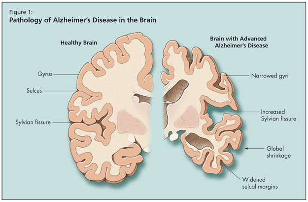
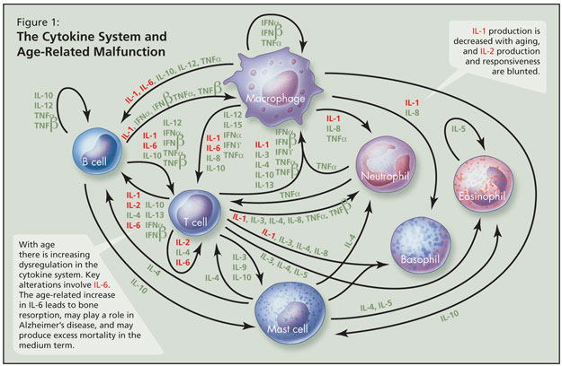
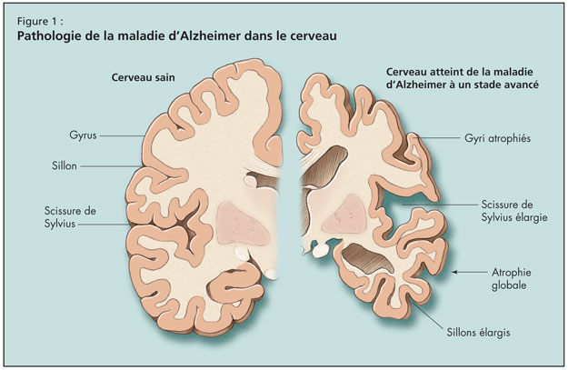
With age there is increasing dysregulation in the cytokine system—a complex series of interacting pro-inflammatory and anti-inflammatory proteins that help to regulate the immune system—and key alterations involve IL-6 (Figure 1). The age-associated increase in IL-6 is a major cytokine derangement, Dr. Alibhai stated, and may parallel chronic inflammation; this leads to bone resorption, may play a role in Alzheimer’s disease, and may produce excess mortality in the medium term. IL-1 production is decreased with aging, and IL-2 production and responsiveness are blunted. He noted that it remains unclear which cytokine changes are key, as no one acts independently. Over time there is more T-cell than B-cell response to infection with age. Not all changes are attributable to aging per se, and subclinical disease and/or environmental factors likely play some role.


The clinical consequence is that older adults are more predisposed to infection but not overt immune deficiency. There is increasing risk of reactivation of dormant infection such as herpes zoster and mycobacterial infection. These risks are compounded by illness and malnutrition. Further, vaccinations are less effective and there is higher risk of parasitic infection.

Treatment options to reverse these aging changes are limited due to a paucity of high-quality, long-term studies, and safety and efficacy of potential therapeutic agents are not clearly established. Early evidence suggests the potential value of melatonin for increased antibody production, helper T-cell activity, and may be able to stimulate IL-2 release. Other data suggest that modified nutritional intake is associated with positive health correlates (e.g., enhanced immunity via increased intake of polyunsaturated fatty acids and vitamin E); further, there has been some benefit shown with modified immunization schedules (e.g., more numerous doses of hepatitis B vaccines in older adults) but larger studies are needed.

The next phase of the presentation concerned changes to the haematological system with age. Generally, concentrations of most blood constituents do not change with age. However, anaemia becomes increasingly common with older age but is not fundamentally age-related (body iron stores increase with age); instead, anaemia may be attributable to disease or other factors (e.g., subtle nutritional deficiencies). Haemoglobin levels may decline slightly with age, although this is still controversial. More importantly, the aged bone marrow cannot quickly respond to acute loss of blood. Thus it takes longer to correct acute anaemia by making new red blood cells. Dr. Alibhai reminded listeners that total white blood cell counts do not change with age, but response is blunted to stresses. Neutrophil and monocyte counts do not change with age, but lymphocyte counts decline slightly. With bacterial infections, there are paradoxical decreases in the number of and proliferative rate of neutrophil precursors, partly due to cytokine dysregulation. Older neutrophils are less able to migrate to the site of injury or generate antimicrobial oxidants in response to attack. Older lymphocytes are more vulnerable to radiation damage.

There may be some hypercoagulability and increased red blood cell viscosity with aging, but data are unclear.

Clinical implications of these haematologic changes include a higher predisposition among older adults to developing and sustaining anaemia in response to bleeding. He reiterated that anaemia is not due to aging and needs investigation. As for dangers of anaemia, he advocated that physicians beware of its association with fatigue, slower recovery after ADL disability, impaired executive function, depressive symptoms, increased hospitalization, and increased mortality. How much is causation versus correlation is an area of ongoing investigation.

Other clinical implications are that in individuals undergoing bone marrow suppression (e.g., with chemotherapy), colony stimulating factors (e.g., granulocyte colony-stimulating factor) are usually recommended for neutropenia prophylaxis in oncology settings.
In closing, he emphasized that most haematological indices do not change with age, and the noninvasive work-up for anaemia (ferritin, B12, RBC, folate, etc.) is equally sensitive and specific as in younger people. Dr. Alibhai drew particular attention to evidence that reticulocytosis is impaired with aging, that reticulocyte response to erythropoietin administration is blunted, and other bone marrow reserves are decreased in response to high demand.

The Aging Liver

The Aging Liver

Teaser: 

David G. Le Couteur, MD, PhD, Professor of Geriatric Medicine, Centre for Education and Research on Ageing and ANZAC Research Institute, University of Sydney and Concord RG Hospital, Sydney, Australia.
Arthur Everitt, PhD, Associate Professor, Centre for Education and Research on Ageing, and Department of Physiology, University of Sydney, Sydney, Australia.
Michel Lebel, PhD, Associate Professor, Centre de Recherche en Cancérologie de l’Université Laval, Hôpital Hôtel-Dieu de Québec, Québec, PQ.

The liver undergoes substantial changes in structure and function in old age. There are age-related changes in liver mass, blood flow, and hepatocyte and sinusoidal cell morphology. These changes are associated with a significant impairment of many hepatic metabolic and detoxification activities, with implications for systemic aging and age-related disease. For example, the age-related impairment of the hepatic metabolism of lipoproteins predisposes to cardiovascular disease. The age-related decline in the hepatic clearance of most medications causes an increased risk of adverse drug reactions. Many of the beneficial effects of caloric restriction and caloric restriction mimetics such as resveratrol are mediated by their effects on the liver. Increasingly, the liver is seen as having a key role in aging.
Key words: liver, aging, hepatocyte, liver sinusoid, drug metabolism.

Managing Abnormal Liver Blood Tests in Older People

Managing Abnormal Liver Blood Tests in Older People

Teaser: 

James Frith, MB ChB, MRCP, Clinical Research Associate, Biomedical Research Centre in Ageing—Liver Theme and Institute for Ageing and Health, Newcastle University, Newcastle, UK.
Julia L. Newton PhD, FRCP, Professor, Biomedical Research Centre in Ageing—Liver Theme and Institute for Ageing and Health, Newcastle University, Newcastle, UK.

The prevalence of chronic liver disease is increasing in older people. The presentation of these diseases is often asymptomatic or nonspecific, so they easily go undiagnosed. Investigating the older person who has abnormal liver function is important in primary care, and the same vigilance should be applied to an older person as to a young person, even with mild abnormalities. Referral for specialist opinion in appropriate older people provides important diagnostic and prognostic information. Treatment options are similar for all age groups. Morbidity and age-adjusted mortality are often more severe in older people; therefore, early diagnosis and intervention are important.
Key words: chronic liver disease, aging, liver function tests.

SOLIDAGE presente un dialogue international : La maladie chronique et le vieillissement

SOLIDAGE presente un dialogue international : La maladie chronique et le vieillissement

Teaser: 

SOLIDAGE présente un dialogue international
La maladie chronique et le vieillissement :
de la recherche aux politiques de santé et à la pratique

SOLIDAGE, le Groupe de recherche Université de Montréal-Université McGill sur la fragilité et le vieillissement, de concert avec FEDERATION CJA, a tenu le 6 novembre 2008 un symposium international sur la maladie chro nique et le vieillissement. Ces deux enjeux clés représentent des défis importants et inextricablement liés auxquels le Québec et les autres systèmes de santé à l’échelle mondiale font face actuellement.

La maladie chronique qui perdure tout au long de la vie a des effets considérables sur le processus de vieillissement, et elle est surtout prévalente dans les dernières années. L’effet cumulatif de la maladie chroniqu e et de la diminution des réserves physiologiques liée à l’âge contribue à l’apparition de la fragilité, de l’incapacité et de la perte d’autonomie chez la population vieillissante. La maladie chronique est devenue le premier facteur d’utilisation des ressources de santé dans notre société. Retarder l’apparition de la fragilité et de la perte d’autonomie servira non seulement à favoriser un vieillissement sain, mais pourra également assurer la durabilité à long terme de systèmes de santé dont le fardeau ne cesse de s’alourdir.

Le symposium international a réuni des décideurs et des responsables de l’élaboration des politiques, des chercheurs, des gestionnaires et des professionnels de la santé, lesquels ont débattu des moyens pouvant & ecirc;tre utilisés par le système de santé du Québec pour bien faire face à ces enjeux, à la lumière de l’expérience internationale et de celle des autres provinces canadiennes.

Commanditaires et partenaires : La maladie chronique et le vieillissement : de la recherche aux politiques de santé et à la pratique

Appuyé par :

Instituts de recherche en santé du Canada

Ministère de la Santé et des Services sociaux Québec

Agence de la santé publique du Canada

Réseau québécois de recherche sur le vieillissement

Ministère de la Famille et des Ainés Québec

Fondation de l’Hôpital général juif Sir Mortimer B. Davis

Hydro Québec

Ministère du Développement économique, de l’Innovation et de l’Exportation Québec

Lawrence S. Bergman, député de D’Arcy-McGee

Pierre Arcand, député de Mont-Royal

Les partenaires :

Hôpital général juif Sir Mortimer B. Davis, Centre d’épidémiologie clinique et de recherche en santé publique, Institut Lady Davis

La Chaire Dr Joseph Kaufmann en gériatrie, Université McGill

Université de Montréal, Département d’administration de la santé,
Groupe de recherche interdisciplinaire en santé (GRIS)

Initiative canadienne sur la fragilité et le vieillissement

Les opinions qui sont exprimées ici ne sont pas nécessairement celles de l'Agence de la santé publique du Canada.