Advertisement

Advertisement

biological and clinical advances

Dementia: Biological and Clinical Advances--Part III

Dementia: Biological and Clinical Advances--Part III

Teaser: 

Christine Oyugi, BSc
Managing Editor,
Geriatrics & Aging.

Edited by:
Karl Farcnik, BSc, MD, FRCPC
Psychiatrist, Division of
Geriatric Psychiatry,
University Toronto,
Part-time staff,
Toronto Western Hospital, Toronto, ON.

Contributions from:
Morris Freedman, MD, FRCPC
Director, Behavioural Neurology
Program, Baycrest Centre for Geriatric Care and Staff Scientist,
Rotman Research Institute, Toronto, ON.

Helena C. Chui, MD
Professor of Neurology,
University of Southern California
Los Angeles, Ranchos Los Amigos
National Rehabilitation Center, Downey, CA, USA.

Ian McKeith, MD, FRCPsych
Professor of Old Age Psychiatry,
Institute for Health of the Elderly
University of Newcastle Upon Tyne, UK.

  • What are the clinical features of Frontotemporal and Lewy Body Dementias?
  • What is the relationship between dementia and vascular disease?
  • How would you differentiate among the different dementias?
  • Does determining the distinction between Mild Cognitive Impairment and dementia have any clinical relevance or is it merely an academic exercise?

These are a few of the topics that were addressed by speakers during the last day of the 11th Annual Rotman Conference. This article summarizes the points presented during the last day of this conference on Tuesday, March 20th.

Dr. Morris Freedman, Director of the Behavioural Neurology Program at Baycrest Centre for Geriatric Care and a Staff Scientist at the Rotman Research Institute in Toronto, provided an extensive clinical review of frontotemporal dementia (FTD). The features of FTD are important for the differential diagnosis of this disease from Alzheimer disease (AD). Patients with FTD have marked personality and emotional changes that include loss of social awareness, and antisocial or disinhibited behaviour (e.g. use of rude speech, neglect of personal hygiene and grooming); they may also become easily distracted. These patients typically have poor insight and may not recognize that they have any behavioural problems. Other features include overeating, excessive smoking, oral exploration of objects and stereotypical behaviour such as wandering.

Language is a key factor in distinguishing FTD from AD. FTD patients have early preservation of language, in contrast to the situation in AD patients where language is primarily affected early in the course of the disease. As AD patients become more impaired, they develop fluent aphasia with comprehension problems. FTD patients experience a reduction in speech capacity, which may eventually lead to mutism, but their comprehension is relatively preserved. Memory loss in FTD is variable and not as severe as that with AD.

The pathophysiology of FTD involves the anterior temporal and frontal lobes. There are two forms of neuropathology--a microvacuolar form and a gliotic form. The microvacuolar form involves the general loss of neurons, microvacuolar degeneration (a spongiform-type of change), a mild astrocytic gliosis and primarily involves laminae I-III. There are no Pick cells or bodies that are seen in Pick's disease, but clinically one cannot differentiate between Pick's disease and FTD. The gliotic form of FTD represents the pathology of Pick's disease and involves all the cortical layers. There is intense astrocytic gliosis and Pick bodies may be present. There has been some debate as to whether the microvacuolar form and the Pick-type pathology represent the same or different disorders.

FTD has an earlier age of onset than does AD; the average age of onset is between 50-60 years of age. The disease duration averages 8-10 years. Although the precise cause of FTD remains unknown, there is a genetic predisposition to the disease in some patients. Fifty percent of patients with FTD have a positive family history and up to 18% have an abnormality on chromosome 17 (autosomal dominant).

There is also a relationship between FTD and motor neuron disease; some patients with FTD also develop motor neuron disease, symptoms of which can appear before or after the onset of FTD. If a diagnosis of FTD is made, it is important for physicians to be aware of the co-occurrence of motor neuron disease.

FTD patients have normal EEG results in the early phases of the disease--in fact an abnormal early EEG argues against a diagnosis of FTD. SPECT analysis shows deficits in frontal and temporal perfusion; however, this is not a diagnostic feature, as AD patients can have the same feature. Neuropsychological assessments show a marked deficit on frontal tests, with an absence of severe amnesia and perceptual 'parietal' deficits (e.g. copy to command is still good).

FTD is one of three prototypical clinical syndromes comprising the broader entity of frontotemporal lobar degeneration: Frontotemporal dementia (FTD), Primary Progressive Aphasia (PPA) and semantic dementia (SD). The difference among the three conditions is based on the location of the pathology--FTD is frontotemporal, SD involves lesions in the temporal lobes bilaterally, and PPA involves left frontotemporal pathology. SD, more so than either FTD or PPA, is easily confused with AD--similar to patients with AD, these patients have fluent speech and comprehension deficits. But unlike those with AD, these patients lose the meaning of words and objects with relatively good preservation of memory. PPA is less likely to be confused with AD because patients' speech becomes non-fluent.

Although patients show serotonergic deficits, there are currently no drugs available for the treatment of FTLD. SSRI's may improve some of the behavioural problems. As patients do not have cholinergic deficits, cholinesterase inhibitors will not help and may actually aggravate symptoms.

Dr. Ian McKeith, Professor of Psychiatry from the Institute of Health in the Elderly in Newcastle, England, updated the conference attendees on the current understanding of Lewy Body Dementia. Lewy body dementia (DLB) accounts for 15-20% of all dementias in old age, but has only been widely recognized since the mid-1990s. The clinical phenotype of Lewy Body Dementia (DLB) is related to the site, severity and amount of Lewy body pathology. DLB is characterized by the presence of Lewy bodies in the brainstem (substantia nigra and locus coeruleus), and in the subcortical (nucleus basalis of Meynert) and cortical regions of the brain. Neuronal loss and gliosis are also present in those areas. In some cases, there is an overlap between DLB, AD and Parkinson's disease (PD). As is the case in patients with both AD and PD, DLB patients have b-amyloid plaques and neurofibrillary tangles in their brains, although not in sufficient numbers to make a diagnosis of AD. The core clinical features of DLB include fluctuating cognitive impairment (seen in 80% of patients), persistent visual hallucinations (70% of patients) and Parkinsonism (75% of patients). These features are used to distinguish between DLB patients and AD patients (See Table 1). Other features which are supportive of DLB but lack specificity are: transient lack of consciousness (40%), falls and syncope (50%), systematized delusion (70%), neuroleptic sensitivity (50%), depression (50%) and REM sleep disorder (no estimates available). DLB is commonly mis-diagnosed as AD, as patients are equally impaired on both the MMSE and the Cambridge Cognitive Examination (CAMCOG). However, DLB patients perform worse on tests of attention (e.g. reaction time), visuospatial performance (e.g. clock drawing) and visual perception (e.g. fragmented letters). A Consensus guideline for the clinical and pathological diagnosis of dementia with Lewy bodies was developed in 1996. Several studies have been performed to validate these criteria and have found that the Consensus criteria for DLB performed as well in prospective studies as did those for AD and vascular dementia (VaD), with a high diagnostic sensitivity. Fluctuation is an important diagnostic indicator, reliable measures of which need to be further developed. Although specificity of the clinical diagnostic criteria is generally high, 17-78% of cases may be missed. This may be attributed to clinicians being unaware of the criteria or unfamiliar with the diagnosis. The potential contribution of neuroimaging to the differential diagnosis of DLB from other dementias remains uncertain, although relative preservation of the hippocampus and temporal lobe is found in DLB when compared with AD.

TABLE 1

Core Clinical Feature of DLB vs. AD

Clinical Feature

DLB

AD

Fluctuating Cognitive Impairment

80%

60%

Persistent Visual Hallucinations

70%

15%

Parkinsonism (bradykinesia, rigidity, gait)

75%

20%

Currently, there is no treatment that stops the progression of DLB. Much of the focus on treatment has been the management of the neuropsychiatric symptoms of the disease and the associated movement disorders. Unfortunately, 50% of patients show sensitivity to older neuroleptics including haloperidol and phenothiazines, and these patients are more likely to die than are those not treated with these drugs. However, newer antipsychotics such as olanzapine and quetiapine may be relatively safer for the management of DLB. Recent studies have shown a benefit of acetylcholinesterase inhibitors with respect to the treatment of behavioural, as well as cognitive, aspects of this disease, and it is possible that these drugs could become the treatment of choice in the future.

Dr. Helena Chui, a Professor from the University of Southern California, gave a talk on cognitive impairment due to subcortical ischemic vascular disease.

Ischemic vascular disease (IVD) is a common cause of dementia in the Western world. Similar to the situation with AD, the incidence of VaD increases with age. However, the exact incidence and prevalence of VaD is difficult to discern. The major problem remains the disagreements with regards to diagnostic criteria and their implementation. In particular, there is uncertainty regarding the following:

  1. The classification of patients who show both vascular and degenerative features (mixed-dementia);
  2. The difficulty choosing among several different clinical criteria (e.g., the Hachinski Ischemic Score);
  3. The use of imaging findings in defining VaD;
  4. The minimal level of disease severity required for a patient to be included in epidemiologic studies.

The problem in diagnosing vascular dementia lies in the causal relationship. It is not very difficult to diagnose dementia and, with the recent advancements in structural imaging, it is also not very difficult to diagnose vascular disease. The conundrum is--what is the relationship between the two and how do we know that the vascular lesion seen in imaging is causing the dementia syndrome? According to Dr. Chui, the term Vascular Dementia is too broad. VaD is not a disease, but only one possible phenotypic expression of vascular brain injury. For this reason, her talk focused on subcortical ischemic vascular dementia (SIVD). There are many types of cerebrovascular disease, leading to variable clinical and symptomatic expressions (Figure 2). There are a number of guidelines available on the effective treatment of risk factors that lead to these conditions and this should be the focus of SIVD management. The frequency of SIVD seems to vary depending on the ethnic group; it is more common in persons of Japanese or African American descent.

The small vessels affected in SIVD are within the brain parenchyma and are small penetrating arterioles approximately 100 to 600 mm in diameter. The predominant risk factors for SIVD are diabetes mellitus, hypertension, amyloid angiopathy (a subset of AD), cerebral autosomal dominant arteriopathy with subcortical infarcts and leukoencephalopathy (CADASIL). SIVD is a term that can be used for either the disease or the dementia syndrome. According to Dr. Chui, SIVD represents a more homogeneous clinical and pathological entity, which may be a more useful target for treatment, especially if the target is the cerebrovascular disease and cerebrovascular brain injury rather than its symptomatic expression.

There are two proposed underlying pathophysiologic mechanisms of how SIVD leads to ischemic brain injury--Occlusion and Hypoperfusion. Occlusion leads to small lacunar complete infarcts, cystic necrosis, and loss of all tissue elements (neurons, axons, glia, astrocytes). It leads to a more homogeneous phenotype including subcortical dementia, affective disorder such as depression, extrapyramidal signs and pure motor and sensory deficits. Hypoperfusion results from widespread narrowing of small penetrating arterioles, leading to incomplete infarction where there is a selective loss of tissue elements. For instance, in the white matter there will first be a loss of oligodendrocytes with demyelination, astrogliosis and, later, a loss of axons. Hypoperfusive ischemic brain injury has been postulated to be the cause of Binswanger syndrome, which is characterized by a combination of deep white matter changes, as well as a slowly progressive subcortical dementia, gait disturbance and urinary incontinence.

Our current understanding of how cognitive impairment relates to SIVD hinges on the lacunar hypothesis. This hypothesis states that the likelihood of dementia is related to the number, size and location of lacunar infarcts within parallel frontal subcortical loops (Prefrontal Cortex-caudate-globus pallidus-thalamus-PFC). However, recent imaging studies showed that the best correlate between dementia and SIVD was atrophy and not the volume or the number of lacunar infarcts. Clinical evaluation of SIVD should include tests of working memory, recognition memory and executive function.

The treatment of SIVD can be divided into three components. The first is primary prevention, where one tries to prevent infarction or vascular cognitive impairment by managing vascular risk factors such as hypertension and diabetes. For secondary prevention, where there is evidence of vascular brain injury--infarction, incomplete infarction or vascular cognitive impairment--the goal is to prevent the recurrence or the progression of disease. There is evidence that, even at this stage, one should continue to manage hypertension but also administer other treatments. Tertiary treatment refers to symptomatic treatment of memory and cognitive impairment. Acetylcholinesterase inhibitors are currently being studied for this purpose; currently, none have been approved for this purpose.

Dr. Chertkow, Associate Professor of Neurology and Neuropsychiatry at McGill University, presented a talk on high and low technological approaches to the early diagnosis of AD. According to Dr. Chertkow, the goal in trying to delineate an early mild cognitively impaired group is to identify which individuals will or will not deteriorate over finite periods of time (5-10yrs). One of the difficulties in studying individuals with Mild Cognitive Impairment (MCI) is the lack of accepted diagnostic criteria. A diagnosis of MCI can be made if patients meet the following criteria: (a) complain of defective memory; (b) normal activities of daily living; (c) normal general cognitive function; (d) abnormal memory function for age; and (e) absence of dementia. However, there are varying inclusion criteria that may overlap with the aforementioned.

Some researchers contend that patients with MCI may be an in-between group (i.e. individuals who are between normal aging and mild AD) (See Dr. Petersen's talk). The issue is to prognosticate and to define those who are going to progress from this state and those who will not. Prognosis in MCI varies depending on how you characterize your group, but severity of symptoms often predicts progression to AD. Researchers are trying to identify biological and cognitive markers that will assist the general physician in delineating MCI individuals who will progress to AD in a finite time period. The characteristic of a good marker is that it should be precise and simple, should be inexpensive, should be reliable and non-invasive and should have the ability to be validated in pathological cases. Recently, Chertkow and colleagues completed a study on mild memory loss in the elderly. The study looked at 90 individuals who passed the above criteria for MCI and followed them for 3-5 years. Over the course of the study, 51 patients deteriorated to dementia (50 of them meeting the criteria for probable AD) and 39 did not deteriorate. Initially, about 15-17% of the MCI individuals progressed to AD each year; however, even after 10 years, approximately 15% of the individuals did not have dementia and did not appear to be progressive. Therefore, there is a subgroup of MCI patients who do not progress to AD.

There were some interesting differences between those individuals that progressed to AD and those that did not. The progressing group had an older age of onset of their symptoms and performed slightly worse on the MMSE at the time of presentation. The researchers further assessed a number of clinical variables--history, risk factors for AD, physical examination--in order to identify predictors for progression of MCI to AD. The only variables useful as predictors were age, the presence of vascular disease, the number of years the individual smoked, the symptom duration and the MMSE score at initial presentation. It was suspected that some of these factors may have been explained by the same variable and a logistic regression analysis was necessary to find out which factors contribute to the prediction. When this was done, the only significant variables remaining were age at onset of memory problems and the MMSE. This predicted progression in about 67% of the MCI group. In addition, retrospectively, individuals who had lack of orientation to time in the MMSE also progressed to AD.

Hippocampal atrophy (MRI volumetrics) may also be useful as a predictor for progression. MCI patients have hippocampal volume that is intermediary between normal individuals and AD patients (who have significant shrinkage). SPECT scanning and APOE genotype did not appear be useful in predicting progression. The researchers set up an algorithm that was a combination of low-tech and high-tech measures--an approach that can be used by physicians in the future. The algorithm allows a physician to establish a score and stratifies the progression of AD in MCI individuals. In the study, individuals that scored zero on the algorithm never progressed to AD and those who scored 4 or more developed AD.

Dr. Petersen, Director of the Mayo Alzheimer's Disease Research Center, gave an update of recent clinical trials on MCI. A definitive diagnosis of AD can only be made after death through the use of neuropathological methods. For the past 15 years, there have been good criteria for probable AD and correspondence between probable AD and definite AD is about 80-90%, if the usual guidelines for diagnosis are used. Research on MCI suggests that there is a transitional point between normal aging and probable AD. The problem for a physician is how to care for a person who presents at this stage and what to tell the family.

The MCI group of patients is an important group to study because they may give us insight into normal aging. From a practical point of view, these individuals may need to be told that they have a cognitive profile that puts them at a greater risk of developing AD, although, as previously mentioned, some patients may not progress to AD. Physicians have to be very careful not to over-diagnose patients with MCI. Do people who fulfil the criteria actually progress to AD at an accelerated rate and, ultimately, can something be done to impede the development of AD in these individuals using cholinesterase inhibitors or secretase inhibitors? Dr. Petersen and colleagues obtained data from the longitudinal study on aging in Rochester, Minnesota. It should be noted that the subjects (largely Caucasian, middle income) were not necessarily representative of the general population. The cognitive function of these subjects has been followed for approximately 20 years. Usually, early in the disease, the patients are not anosagnosic but are actually aware of their memory impairment. MCI patients that meet this criteria progress to probable AD at a rate of 12% per year compared to controls that progress at a rate of about 1-2% per year. According to Dr Petersen, after 10 years, 80% of these patients progress to AD. There are qualitative features that help predict who is more likely to progress to AD and who is not. The inability of persons to benefit from cues, and hippocampal atrophy, were positive predictors of progression.

Currently, clinical trials are underway to test the use of all the second-generation cholinesterase inhibitors in MCI (Table 2) as well as vitamin E, and COX-2 inhibitors. MCI individuals, if well characterized, present a sample population that will progress to AD at a known rate and are an important target group for preventive therapy. Finding a control for this study group is difficult--age-appropriate controls could be contaminated, as they may include subjects who themselves have MCI.

TABLE 2

Clinical Trials in MCI

Sponsor

Duration of Study

Endpoint

Drugs being tested

Alzheimer's Disease Cooperative Study (ADCS)

3 years

Clinical probable AD

Vitamin E
Donepezil

Merck Frosst

2-3 years

Clinical probable AD

Rofecoxib

Novartis

2 years

Clinical probable AD

Rivastigimine

Janssen-Ortho

2 years

Clinical probable AD

Galantamine

Pfizer

6 months

Symptomatic improvement

Donepezil

Most individuals with MCI will go on to develop AD. In the future, we may also determine predictive phases of other dementias, where a patient can present with slight impairment in multiple domains, or a slight impairment in a single, non-memory domain. These could be used as predictors of the development and progression of several conditions including Frontotemporal dementia, Lewy body dementia, or even primary progressive aphasia.

At least for the relationship between MCI and AD, we now have available criteria that can allow for clinical trials to determine the efficacy of intervention at this stage, possibly preventing the inevitable progression toward AD.

Dementia: Biological and Clinical Advances--Part I

Dementia: Biological and Clinical Advances--Part II

Further Readings

  1. Barber R, Ballard C, McKeith IG, Gholkar A, O'Brien JT, Volumetric study of dementia with Lewy bodies--A comparison with AD and vascular dementia Neurology 2000;54:1304-1309.
  2. Chui H. Dementia due to subcortical ischemic vascular disease. Clin Cornerstone 2001;3(4):40-51.
  3. Jack CR Jr, Petersen RC, Xu Y, et al. Rates of hippocampal atrophy correlate with change in clinical status in aging and AD. Neurology. 2000 Aug 22;55(4):484-89.
  4. Longstreth WT Jr, Manolio TA, Arnold A, Burke GL, Bryan N, Jungreis CA, et al. Clinical correlates of white matter findings on cranial magnetic resonance imaging of 3301 elderly people. The Cardiovascular Health Study. Stroke 1996 Aug;27(8):1274-82.
  5. McKeith IG, Ballard CG, Perry RH, Ince PG, O'Brien JT, et al. Prospective validation of consensus criteria for the diagnosis of dementia with Lewy bodies. Neurology. 2000 Mar 14;54(5):1050-8.
  6. Petersen RC, Stevens JC, Ganguli M, Tangalos EG, Cummings JL, DeKosky ST. Practice parameter: early detection of dementia: mild cognitive impairment (an evidence-based review). Report of the Quality Standards Subcommittee of the American Academy of Neurology.Neurology. 2001 May 8;56(9):1133-42.
  7. Petersen RC. Aging, mild cognitive impairment, and Alzheimer's disease. Neurol Clin. 2000 Nov;18(4):789-806. Review.
  8. Petersen RC, Smith GE, Waring SC, Ivnik RJ, Kokmen E, Tangelos EG. Aging, memory, and mild cognitive impairment. Int Psychogeriatr 1997;9 Suppl 1:65-9.
  9. Rocca WA, Kokmen E. Frequency and distribution of vascular dementia.Alzheimer Dis Assoc Disord 1999 Oct-Dec;13 Suppl 3:S9-14.
  10. Shah S, Tangalos EG, Petersen RC. Mild cognitive impairment. When is it a precursor to Alzheimer's disease? Geriatrics. 2000 Sep;55(9):62, 65-8. Review.

Dementia: Biological and Clinical Advances--Part II

Dementia: Biological and Clinical Advances--Part II

Teaser: 

Christine Oyugi, BSc
Managing Editor,
Geriatrics & Aging.


Edited by:
Karl Farcnik, BSc, MD, FRCPC
Psychiatrist, Division of Geriatric
Psychiatry, University Toronto,
Part-time staff,
Toronto Western Hospital, Toronto, ON.

Contributions from:
Serge Gauthier, MD, FRCPC
Director, McGill Centre for Studies in Aging,
Professor of Medicine (Neurology),
McGill University, Montreal, QC.


Dr. Katarina Rogaeva
Research Associate, Centre for
Research in Neurodegenerative diseases,
University of Toronto, Toronto, ON.

Introduction
With Canada's aging population, dementia has become a growing problem. Eight percent of Canadians who are over the age of 65 suffer from dementia, of these approximately 60% are believed to have Alzheimer disease. Dementia is an age-related disease, with the prevalence increasing from 2.4% of those from 65-74 years of age, to 34.5% of those 85 and older. There are sixty thousand new cases of dementia diagnosed each year, and the costs of providing health care for these patients continue to escalate. It is with these alarming statistics in mind that clinicians gathered to hear the latest developments in the biology of dementias presented during the 11th annual Rotman conference. Various dementias were discussed at the conference including Frontotemporal lobar dementia (FTLD). FTLD is the third most common form of cortical dementia following Alzheimer disease (AD) and Dementia with Lewy Bodies. It is often mistaken for AD, yet it presents strikingly different clinical and histopathological features and therefore, must be managed distinctly. This article will serve as a summary of some of the key points presented during the first day of this conference on Monday, March 19th.

In her opening remarks, Dr. Rogaeva, a Research Associate at the Centre for Research in Neurodegenerative diseases at the University of Toronto, gave a brief overview of the genetic variability and pathobiology of Alzheimer Disease. AD is a neurological disorder that is characterized by a slow degenerative process affecting cognitive function. At a histopathological level, AD patients are characterized by the deposition of senile plaques within the brain, as well as within the walls of cerebral blood vessels. It is believed that through some unknown mechanism, these senile plaques exert a toxic effect on surrounding neurons, resulting in the neuronal degeneration found in AD patients. Other pathology that is seen includes reactive microglia, swollen neurons and intracellular neurofibrillary tangles.

The primary constituent of these senile plaques is the amyloid b peptide (Ab). Two proteases, b-secretase and g-secretase cleave this peptide from a larger precursor protein, b-amyloid precursor protein (b-APP). Essentially, b-secretase cleaves APP to produce an APPsb soluble fragment.

The pathogenesis of AD has been linked to both genetic and environmental factors (See Figure 1).

A number of genes have been identified as influencing the development of AD including APO E, the presenilin genes and b-APP gene. (See Figure 2).

Of these, presenilin 1 (PS1) mutations are said to account for majority of the early-onset familial AD cases. In a study that screened AD patients for PS1 mutations, 11% of the cases had mutations in regions coded by PS1 and, of these, 21 were novel mutations. The study also found that the AD patients who had a positive PS1 test were significantly younger (46 ±11yrs) than were the patients who tested negative for PS1 (60±11yrs).

According to Dr. Rogaeva, screening for presenilin mutations is likely to be successful and cost-efficient if it is targeted to the right groups.

The deposition of Ab in the brains of AD patients is linked to the pathology that is characteristic of the disease. In his talk, Dr. Younkin, Director of Research and Professor of Pharmacology at the Mayo clinic in Jacksonville, explained the role of Ab aggregation in the pathogenesis of AD with a focus on whether it is an essential early event in the disease process. One clue to the role of Ab can be garnered from patients with Down Syndrome or trisomy of chromosome 21. These patients invariably develop AD pathology by age 40 and have been found to have high Ab levels in their plasma. Interestingly, this increase in plasma levels of Ab is seen prior to the onset of symptoms. Studies have shown that all mutations that are linked to AD increase the extracellular concentrations of Ab; this phenomenon occurs prior to the development of the disease and fosters Ab aggregation. Aggregated Ab has been shown to be directly toxic to neurons in culture; for this reason inhibition of b or g-secretase, thereby reducing Ab concentrations, has emerged as a therapeutic target for AD. Clinical trials are currently underway to assess the efficacy of immunization with b-secretase.

Plasma and cerebrospinal fluid (CSF) levels of Ab have been shown to increase with age (over age 65 years). A study comparing patients with typical late-onset AD to age-matched controls found that Ab was increased in AD patients. Some of these patients had levels similar to those found in patients with trisomy 21 or familial AD. What is interesting with late onset AD is that, as the disease progresses, there is a decline in the levels of Ab in the plasma and CSF. The reason for this decline is not well understood, although microglial clearance has been implicated.

Although plasma levels of Ab are not adequate for making a diagnosis of AD, they may be useful as a biomarker for the disease. According to Dr Younkin, in the future, therapies could be targeted to patients with elevated Ab in the same way that patients are treated for elevated cholesterol levels to prevent cardiovascular disease. The study of Ab is only one of many approaches to the study of AD. Other approaches could play important roles in the disease progression and could also be key targets for therapeutic intervention.

Dr. Wilhelmsen, Associate Professor of Neurology at the University of Carlifornia, gave a brief overview on the pathobiology of taupathies. His talk was based on clinical evidence gained from a family with members that suffered from a variety of neurodegenerative diseases. Some of the symptoms observed included: behavioural disinhibition, dementia--which differed from that seen in AD in that there was a relative preservation of language and praxis--and Parkinson's disease, without the typical tremor and non-responsive to L-DOPA. Most members of the family with the disease eventually developed amyotrophy--which is the loss of motor neurons resulting in the development of brisk reflexes, but reduced motor power in the limbs. However, by the time that most patients were dying they were all akinetic, in a fetal position and bed-ridden, making it difficult to recognize that they had typical signs of motor neuron disease. Personality changes were observed in family members including aggressiveness, depression, alcoholism, hyper- and hypo-sexuality, childishness, craving for sweets and hyper-religiosity. Interestingly, they did not respond very well to neuroleptics. The age of onset of symptoms ranged from 25-56 years.

At the time of autopsy, microvascular changes were observed in the anterior temporal cortex of the frontal lobes, between layers II and III. Other pathology includes, rare ballooned neurons, swollen vacuolated anterior horn cells, loss of pigmented cells in the substantia nigra and gliosis in the hippocampus.

Linkage analysis was performed and a link was found to chromosome 17. This analysis was done on several families all of whom had suffered from a variety of disorders including progressive Parkinsonism and dementia with palido-ponto-nigral degeneration (PPD), frontotemporal dementia and primary progressive aphasia (PPA). Several of these families had mutations in the tau gene, which has been implicated in the formation of neurofibrillary tangles in AD. Tau protein deposits have been linked to a variety of neurodegenerative diseases, many of which are frontotemporal dementias or movement disorders, collectively referred to as tauopathies--Pick's disease, progressive supranuclear palsy, and corticobasal degeneration.

What is the function of tau? The tau gene is large and has a complex pattern of alternative spicing. The protein has domains that have been shown to affect the assembly of microtubules. Disruption of this alternative splicing is enough to cause a dysfunction of the tau protein and resulting neurodegeneration. It appears that the regulation of splicing of the gene is important for maintenance of normal brain function. Future research is needed to elucidate whether it is the aggregation of the protein that ultimately results in disease.

Many doctors have never seen a case of progressive supranuclear palsy (PSP). The disease is often misdiagnosed as Parkinson's disease until the patient fails to respond to normal therapy for PD. The prevalence of the disease is less than that for PD. The pathology of PSP is marked by the precipitation of tau protein throughout the midbrain. Although PSP is primarily a sporadic disease, studies show that a precipitating mutation may be required for the disease to occur.

Aggregates of tau protein are seen in many neurodegenerative disorders suggesting that this process may be a common pathway in the pathology of these diseases. Although the importance of the tau gene is known, more research is needed into the interaction of tau with other genes, as well as the regulation and metabolism of tau. Future efforts to develop animal models of tau-mediated neurodegeneration should provide further insights into the onset and progression of tauopathies, as well as Alzheimer disease. This could lead to the discovery of effective therapies for these disorders

The next speaker was Dr. David Westaway who began his review of the latest research in prion diseases. Prion diseases, or transmissible spongiform encephalopathies, are fatal neurodegenerative disorders. These neurodegenerative diseases include scrapie in sheep, mad cow disease in cattle, and Creutzfeldt-Jakob disease (CJD) in humans. Prion diseases may present as genetic, infectious or sporadic disorders, all of which involve the post-translational modification of the prion protein (PrPC) into the disease-causing PrPSc. CJD generally presents as progressive dementia where as the other forms of prion disease typically manifest as ataxia-like disease.

More than 20 mutations of the PrPC gene are now known to cause the inherited human prion diseases, and significant genetic linkage has been established for five of these mutations. However, why or how these mutations cause the protein to change and result in disease is not really understood.

The function of PrPC is still unknown, although there is some evidence that it is involved in the binding of Cu(II) ions. Recent studies suggest that PrPC may function in signal transduction through a pathway involving Fyn tyrosine kinase. Fyn is associated with acetylcholine receptors in muscle cells. It is highly expressed in the limbic system and may have a role in myelination. It was hoped that deletion of the mouse Prnp gene would alter the phenotype such that conclusions could be made with respect to the function of PrPC. Studies in mice show that injection with PrPSc results in death within 150 days. However, Prnp0/0 knockout mice are resistant to prion infection. This provides evidence that cellular PrP is necessary for disease pathogenesis.

Dr. Sandra Black then gave an overview of the use of neuroimaging biomarkers for AD. An ideal test for AD should: reflect the pathophysiology of the disease, allow for pathologic evaluation, allow for early detection, differentiate AD from other dementias, be non-invasive, be affordable, have a sensitivity greater than 80% for detecting AD and a specificity of greater than 80% for distinguishing other dementias. The desired sensitivity and specificity of a biomarker depends on its purpose.

Dr. Black suggested that current clinical techniques can be termed the "bronze standard" for diagnosis. This is based on the combined use of history, and physical and cognitive examination, as well as blood test and neuroimaging to exclude secondary causes. The "gold standard" is based on tissue pathology. Biochemical markers for AD include apolipoprotein E (ApoE) genotyping, and several potential CSF markers including: beta-amyloid, possibly reflecting amyloid deposition and formation of senile plaques; PHFtau protein as a marker for the phosphorylation state of tau, and formation of neurofibrillary tangles; (total) tau protein, a normal axonal protein, used as a marker for ongoing neuronal and axonal degeneration, and the CSF/serum albumin ratio, as a marker for blood-brain barrier damage, used to exclude patients who also have cerebrovascular pathology.

Functional imaging techniques such as PET and SPECT also serve as important diagnostic tools. Recently, functional studies have shown abnormalities in the posterior cingulate and medial temporal regions of patients who show memory impairment and later develop AD. This finding is potentially useful in detecting pre-clinical AD, but it is difficult to apply to individual cases.

With the emerging therapeutic compounds for the treatment of AD, an important role of imaging is in monitoring whether treatment is actually slowing down the progression of atrophy. Several techniques have been proposed to monitor the progression of the atrophy, but currently, none can be performed reliably. It is likely that both quantitative neuroimaging and biochemical profiles (urine and serum) together with clinical neurobehavioural assessments will be needed in order to achieve the required sensitivity for diagnosing and monitoring AD. Dr. Black stressed the urgent need for sensitive and specific tests.

Dr.Serge Gauthier gave the final talk of the day on currently available and future therapies for AD and related conditions. In his opening remarks, Dr. Gauthier stressed that it is important for clinicians to be knowledgeable of the fact that AD is not just one condition, but entails a mild stage AD, moderate AD and severe AD. Different treatments are effective at different stages in the disease.

In typical cases, AD progresses through relatively predictable stages, as described in the Global Deterioration Scale of Reisberg et al., (Reisberg B, Ferris SH, Deleon MJ, Crook T. The global deterioration scale for assessment of primary degenerative dementia. Am J Psychiatry 1994;44:2203-6). The estimated time from diagnosis to death is usually 5-8 years. In typical AD, most patients initially present with a change of mood, which improves over time. They then experience a linear cognitive and functional decline including a loss of autonomy for instrumental and self-care activities of daily living. Most patients have some degree of neuropsychiatric change, including hallucinations, misidentifications (Capgras syndrome), delusions and paranoid ideation, aggression or apathy, wandering and sexual disinhibition. The appearance of early-onset motor or gait disturbances, including asymmetrical grasp responses is atypical and suggests conditions other than AD. Dr. Gauthier described milestone steps in the progression of AD. These are important as they may act as future therapeutic targets (Table 1).

TABLE 1

Milestones in Progression of AD

a) Diagnosable dementia
b) Loss of IADL (Instrumental Activities of Daily Living)
c) Emergence of neuropsychiatric symptoms
d) Nursing home placement
e) Loss of basic ADL
f) Death

Different stages of AD present different issues in management of the disease. In the early stages of AD the management issues include making an accurate diagnosis, patient and caregiver education, advance power of attorney, advance directives and whether the patient should continue driving. In the moderate stages, the physician should carefully monitor the patient's autonomy. At this stage it is very important to monitor the health and well-being of the caregiver. Management issues in the final, severe stages of AD include cessation of cholinesterase inhibitors and end-of-life decision making.

Taken together the speakers presented a great deal of useful information on the pathobiology of a variety of neurodegenerative diseases. Understanding the genetic and biochemical basis of these diseases may allow for treatments tailored to various disease stages, as well as providing useful biomarkers to enable detection of those patients who are most at risk.

Dementia: Biological and Clinical Advances--Part I

Dementia: Biological and Clinical Advances--Part III

Dementia: Biological and Clinical Advances--Part I

Dementia: Biological and Clinical Advances--Part I

Teaser: 


Cognitive Assessment and Neuroimaging of Dementia

Bob Chaudhuri, MD
Department of Psychiatry,
University of Toronto,
Toronto, ON.

Contributions from:

Morris Freedman, MD, FRCPC
Director, Behavioural Neurology
and Senior Scientist,
Rotman Research Institute,
Baycrest Centre for Geriatric Care,
Professor of Medicine (Neurology),
University of Toronto,
Toronto, ON.

Larry Leach, PhD, CPsych
Psychologist, Department of Psychology,
Baycrest Centre for Geriatric Care,
Adjunct Professor,
University of Toronto,
Toronto, ON.

Wendy Meschino, MD, CCFP, FRCPC, FCCMG
Clinical Geneticist,
North York General Hospital,
Toronto, ON.

On Sunday, March 18th, a series of speakers discussed cognitive assessment and neuroimaging in dementia. The speakers included Dr. Morris Freedman, Dr. Larry Leach, Dr. Robert van Reekum, Dr. Sandra E. Black and Dr. Wendy Meschino.

The focus of Dr. Freedman's workshop was the Bedside Assessment of Cognitive Function. He demonstrated cognitive screening tools in dementia and selective supplementary testing from the Behavioural Neurology Assessment that was developed at Baycrest and that is currently being prepared for publication.

Dr. Freedman showed how clock drawing is an easy to administer and sensitive measure of cognitive function. However, not all time settings are equally good for demonstrating deficits. A preferred time to ask the patient to draw is "10 after 11." He gave striking examples in which the clock-drawing test was more sensitive than the Folstein Mini-Mental Status Exam (MMSE). He pointed out that, in conjunction with the clock drawing, mental status testing in the office setting can be effective in defining and tracking the patient's cognitive function.

There are a variety of common clinical problems that are associated with making a diagnosis of dementia.

The first problem is how best to test the patient's memory. Memory is almost always impaired in Alzheimer disease, although poor memory does not necessarily mean AD; intact memory suggests a diagnosis other than AD.

Generally, a patient has a history of memory loss and he or she, and/or the family, is concerned about the possibility of dementia.

The second problem is how best to test the patient's attention. The MMSE uses serial 7s and spelling the word 'world' backwards as tests; however, using months backwards or, in severe cases, having the patient use serial 1s subtraction from one hundred is useful.

A third problem is how to assess language. Dr. Freedman stated that listening to the pattern of spontaneous speech (fluent vs. nonfluent), and testing comprehension, naming and repetition are very useful. Fluent speech suggests a temporal-parietal lesion, and non-fluent points to a frontal lesion. AD produces fluent speech until the later stages.

Auditory comprehension testing involves the assessment of single words, phrases and whole body commands. He suggested that the clinicians also test repetition of single words and phrases, and test naming by showing the patient common objects to name (e.g. pencil and watch). In Alzheimer disease, naming is impaired and repetition is normal in the early stages.

Dr. Freedman outlined testing for ideomotor apraxia, which is the inability to pretend to carry out a motor activity to command (e.g. comb your hair) when the activity is one that can be performed easily in spontaneous situations. To determine if a patient has apraxia, give a verbal command. If the patient fails to respond correctly to the command ask him or her to imitate the action. Finally, ask the patient to use the real object if imitation is impaired.

Visuospatial function is commonly impaired early in the course of AD. Clock drawing is a sensitive test of visuospatial function. Supplementary testing includes drawing to command and copy, such as a house, a flower and a cube.

Dr. Freedman summarized by stating that there are a number of tests of memory. These include asking patients the year, month, day, place, name of the Prime Minister and Premier and immediate and 5 minute 3 word recall words such as cat, apple, table. Attention can be tested through serial 7s and 3s subtraction and by reciting the months backwards. Naming can be tested by asking patients to name objects. Asking the patient to draw a clock, with the time set to 10 after 11, is an effective method to test visuospatial function. Testing similarities and proverb interpretation assesses the ability to manipulate acquired knowledge. It should be noted that the tests of similarities and proverbs may be somewhat confounded by cultural biases. Copying patterns of multiple loops or alternating sequences can be used to assess frontal lobe function. Perseveration on these tasks, and deficits on world list generation of animal names or words beginning with the letter F, are often seen following frontal brain damage.

This workshop reviewed the basic aspects of the mental status exam that make up screening assessment in an office setting. Dr. Freedman concluded that testing cognitive function is very important for the differential diagnosis of dementia.

Dr. Larry Leach presented the next workshop on Neuropsychological Assessment in Dementia. The learning objectives of this lecture were: to determine whether an individual met the basic criteria for dementia; to evaluate the effectiveness of cognitive testing in identifying dementia; and to establish a battery of tests that describes the cognitive profile of a patient with dementia.

The DSM-IV modified definition of dementia was discussed and compared to the definitions provided by Cummings and Benson (1983) and by Strub and Black (1981).1,2

The test battery domains were discussed in terms of memory, abstract reasoning, perceptual functioning, constructional ability, language, praxis, mood, global intelligence and cognitive functioning.

Common referral questions include: a) "Is impairment present?" b)"What is the pattern of impairment?" c)"What is the etiology?" d) "Is it mood related?" d) "And has there been a change?"

Dr. Leach also discussed diagnostic issues involving the effects of age, education, gender and culture.

The Prevalence of dementia was discussed and found to have an incidence of 2.4% in the population aged 5 to 74, 11.1 % in the population aged 75 to 84, and 34.5% in the population over age 85.

Dr. Leach reached several conclusions regarding the use of the MMSE as a diagnostic tool, including that:

a) It was poor for diagnosing dementia when prevalence is less than .60%;

b) It was adequate for ruling out moderate to severe cognitive impairment when prevalence is below .35%, except for those patients who are over the age of 80 and had a lower educational status. Cut-off scores need to be adjusted according to age and education.

The following criteria for diagnosing 1) Frontal-Temporal Dementia, 2) Lewy-body disease, 3) Vascular Dementia, and 4) Alzheimer Dementia, were discussed. Special tests for dementia were discussed.

Cognitive and neuropsychological tests provide insights into the nature and severity of brain dysfunction as well as brain regions that are dysfunctional in dementia. The pattern of impairment reflects brain regions affected more so than the cause of dysfunction. Therefore, there are practical limitations to diagnosing cause based solely or primarily on the results of mental status examination or neuropsychological assessment. Despite this limitation, the pattern of deficits due to dementia are clearly distinguishable for those cognitive disruptions associated with depression.

Dr. Robert van Reekum followed Dr. Leach with a presentation on the importance of neuropsychiatric evaluation in dementia. He emphasized that changes in mood and behaviour are common in these patients and can cause suffering, impact on disability and handicap, influence diagnosis and have prognostic implications. Perhaps most importantly, these changes in mood and behaviour are treatable. He divided factors that should be assessed into premorbid factors, which included a past history of medical, psychiatric, personal, neurodevelopmental and social factors, and responses to previous treatments, and current factors. The current factors include medical status, arousal antecedents/precipitants/patterns, and cognitive and neurologic status.

A variety of different behaviours are common in patients with dementia. Because the actual dementia may mimic or mask psychiatric disorders, the evaluation of psychiatric illness in this population must take into account the direct effects of CNS disease. Some of the behavioural symptoms of dementia, such as psychosis, may warrant pharmacological intervention. Anxiety, agression, disinhibition and apathy may also warrant treatment.

Finally, Dr. van Reekum stressed the need for structure, reliable and valid Behavioural inventories to improve the consistency of behaviour quantification in these patients. He reviewed one such inventory, the Neuropsychiatric Inventory (NPI).

Dr. Sandra Black presented the pros and cons of the currently available techniques for neuroimaging in dementia. The objectives of this workshop were:

a) To review currently available structural and functional imaging techniques;

b) To review principles for the interpretation of brain-behavior relationships in dementia;

c) To illustrate the above in case examples of patients with dementia.
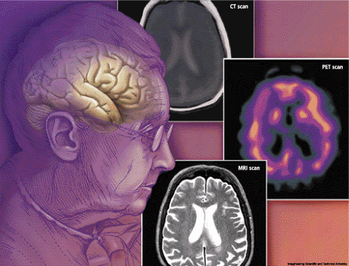
dementia image

Currently available techniques for neuroimaging in dementia include magnetic resonance imaging (MRI), positron emission tomography (PET) and CT scan. The relative merits of these techniques were discussed.

CT scanning in dementia has the following strengths: it has excellent spatial resolution; it is relatively cheap and widely available, it rules out major pathologies; and, if applied correctly, medial temp width can be measured. However, it also presents the following weaknesses: there is less contrast; there is a problem of bone artifact; it is less sensitive to pathology; and the patient is exposed to radiation.

Magnetic resonance imaging (MRI) gives excellent spatial resolution, no bone artifact and better contrast with high sensitivity, and is low risk. However, it has a higher cost than does CT scanning and is less readily available and takes longer to image (but this is changing), and there are associated contraindications (pacemaker, aneurysm and claustrophobia).

Positron Emission Tomography (PET) has the strength of being versatile: injected radiolabel can measure regional cerebral blood flow, metabolism or receptors; direct quantification is possible; there is fair resolution; and it can measure brain activation using subtraction. On the downside, it has a high costs associated with it (cyclotron and special team); it is a scarce resource and not widely available; and there is a risk associated with exposure to radiation.

Single Photon Emission Computed Tomography is relatively cheap and widely available, and the injected radio label measures regional brain perfusion; but it only offers relative quantification, gives poor spatial resolution and again has a risk of radiation exposure.

All of these techniques are useful in different ways for the diagnosis of dementia. Examination of blood flow, oxygen utilization, cerebral atrophy and brain function can be demonstrated using these techniques.

The final lecturer was Dr. Wendy Meschino who discussed the use of Genetic Testing for Dementia in Clinical practice. The objectives of the presentation were how to approach a family history of dementia, risk assessment of hereditary dementia, what tests are available, determining when testing is helpful and providing information on how to get testing done. Alzheimer disease risk factors were identified as: increasing age, a positive family history of AD, Down Syndrome, cognitive impairment, head injury, low education level, and aluminum exposure (controversial). Exposure to exogenous estrogen for women and the presence of arthritis may be protective.

The family history of dementia work-up includes:

a) Taking a detailed three-generation pedigree, noting specific symptoms, such as the age of onset and the number of unaffected relatives;

b) Obtaining medical records, including autopsy, to determine whether the patient suffers from AD or some other condition.

Less than 5% of AD is inherited as an autosomal dominant trait. These cases are usually early in onset. Hereditary factors combined with environmental factors (complex inheritance) play a role in a further 15-25% of mostly late-onset cases. The remaining 75% are sporadic, late-onset and indistinguishable in phenotype from hereditary forms.

Dr. Meschino reviewed a list of genes that are now known to cause hereditary dementias. For early-onset AD these include Presenilin 1, APP and Presenilin 2. Presenilin 1, a gene on chromosome 14, accounts for the majority of early-onset cases. The average age of onset is in the 40's. Some cases of frontotemporal dementia (FTD) are associated with mutations in the tau gene. Notch3 mutations have been found in patients with CADASIL (cerebral autosomal dominant arteriopathy with subcortical infarcts and leukoencephalopathy).

There are also a number of genes that are believed to predispose a patient to late-onset AD. These risk modifier genes are: the e4 allele of the APOE gene on Chromosome 19 (e4 has the effect of decreasing the age of onset), and A2M-2 on Chromosome 12 (in some studies associated with an increased risk of developing AD). These are examples of genes which affect susceptibility to disease, but do not directly cause it. Genetic testing for APOE is not recommended for asymptomatic individuals because the test cannot determine whether an individual will or will not develop AD in the future.

Prior to genetic testing, patients should be provided with genetic counselling, especially when undergoing pre-symptomatic testing. Important information to cover in the session includes outlining the differences between hereditary and sporadic AD, late-onset and early-onset AD, and the risk of developing AD in the general population compared to the risk for that individual.

There are a number of important ethical issues that need to be considered when discussing predictive testing. The patient must be able to make an informed choice; that is, there should be no patient coercion. The clinician should outline the various reasons for knowing or not knowing the diagnosis, the effects it may have on the family and the potential that they will be subject to discrimination. In general, requests for prenatal diagnosis for adult-onset diseases are infrequent, and testing in childhood is strongly discouraged.

One of the following criteria should be met before Alzheimer testing is considered:

a) An individual affected with AD, with onset at less than 60 years;

b) A first-degree, unaffected relative of an affected individual, in a family with 2 or more early-onset cases (all affected are deceased);

c) 2 or more living affected family members with onset greater than 60 years (DNA samples needed from both relatives).

As part of this presentation a video clip was shown from a recent CBC Nature of Things episode called Amanda's Choice, in which a young woman from northern Ontario underwent genetic testing for early-onset Alzheimer disease. There was an extensive family history of the disease in her mother's family with onset of the disease in the mid-30's. She was shown receiving her genetic test results from the presenter as well as genetic counselling. The film explored the emotional impact of living at risk for this devastating disease and the effects on her family.

In summary, these workshops were highly educational and practical. From the neuropsychiatric assessments, MRI and PET diagnostic tests for different dementias, to the ethics and practicality of genetic testing, these workshops appealed to the novice and expert alike.

Dementia: Biological and Clinical Advances--Part II

Dementia: Biological and Clinical Advances--Part III

References

  1. Cummings JL, Benson DF. Dementia: A Clinical Approach 1983. Butterworths & Company, Canada.
  2. Strub RL, Black, FW. The Mental Status Examination in Neurology 1981. Philadelphia: FA Davis.