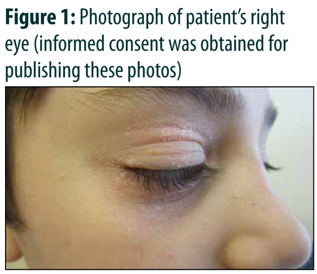
Advertisement

Advertisement

pediatric

Pediatric Psoriasis

Teaser: 

Yvonne Deng,1 Joseph M. Lam, MD, FRCPC,2

1 Faculty of Medicine, University of British Columbia, Vancouver, British Columbia, Canada.
2Department of Pediatrics, Department of Dermatology and Skin Sciences, University of British Columbia, Vancouver, British Columbia, Canada.

CLINICAL TOOLS

Abstract: Psoriasis is a chronic inflammatory skin disease that affects up to 1.4% of children (aged <18 years) with a strong genetic predisposition and is mediated by dysregulation in the crosstalk between the innate and adaptive immune responses. It can have significant impact on quality of life for many patients. There are various subtypes of psoriasis with plaque psoriasis being the most common presentation in both adults and children. Pediatric psoriasis is primarily a clinical diagnosis. With the advent of biologics, the treatment landscape for pediatric psoriasis has shifted and encompasses diverse modalities of therapeutics, including topical and systemic treatments, as well as phototherapy.
Key Words: pediatric psoriasis, psoriasis, chronic, inflammatory, skin disease.

Members of the College of Family Physicians of Canada may claim MAINPRO-M2 Credits for this unaccredited educational program.

www.cfpc.ca/Mainpro_M2

You can take quizzes without subscribing; however, your results will not be stored. Subscribers will have access to their quiz results for future reference.

Psoriasis affects the pediatric population and is associated with negative effects on quality of life and psychological impairments.
Lesions of pediatric plaque psoriasis may be thinner, smaller, more macerated than those classically in adult and present more commonly on the scalp, face, intertriginous areas, and extremities (flexural surfaces for younger children and extensor surfaces for older children).
Psoriasis is not an isolated condition and is correlated with higher rates of myocardial infarction, diabetes mellitus, hypertension, obesity, arthritis and liver disease.
In mild to moderate cases of psoriasis, topical therapies with a short course of corticosteroids and/or vitamin D analogue are first-line. In moderate to severe cases, narrowband UVB phototherapy, systemic agents, such as methotrexate, and biologics should be considered.
Pediatric patient should be assessed for risk factors for associated comorbidities.
A history of preceding streptococcal or viral infection can be suggestive of guttate psoriasis, which is more common in children than adults.
Approach to treatment should be guided by the extent and severity of disease, which can be quantified by BSA, the PASI score and quality of life index surveys.
To have access to full article that these tools were developed for, please subscribe. The cost to subscribe is $80 USD per year and you will gain full access to all the premium content on www.healthplexus.net, an educational portal, that hosts 1000s of clinical reviews, case studies, educational visual aids and more as well as within the mobile app.
Disclaimer: 
Disclaimer at the end of each page

The Non-Operative Management of Scoliosis

Teaser: 

Brett Rocos, BSc (Hons), MB ChB, MD, FRCS (Tr & Orth),

Paediatric Spine Fellow, The Hospital for Sick Children, Toronto, ON.

CLINICAL TOOLS

Abstract:Scoliosis is a common condition that every primary care provider will encounter. There are many treatments available in its management, including observation, physical therapy, pain management strategies, casting, bracing and surgery. In this narrative review, the roles of each of the non-operative strategies in managing adult and paediatric scoliosis are explored, and the evidence supporting each is summarised. Scoliosis affects people at every stage of life, and an understanding of the treatments available will aid in counselling patients and making appropriate referrals.
Key Words: Scoliosis, conservative, paediatric, bracing, physiotherapy, alternative therapies, spine cast.

Members of the College of Family Physicians of Canada may claim MAINPRO-M2 Credits for this unaccredited educational program.

www.cfpc.ca/Mainpro_M2

You can take quizzes without subscribing; however, your results will not be stored. Subscribers will have access to their quiz results for future reference.

• Scoliosis is common
• Most patients require observation only
• Patient information is essential
• Casting and bracing have roles in the growing skeleton only
• Physical therapy has limited evidence in both adult and paediatric deformity
• Alternative therapies have no proven use in the management of scoliosis
The majority of patients with scoliosis can be observed
Reliable patient information is critical
There is limited evidence that physiotherapy is effective, and no evidence that alternative therapies are effective in treating scoliosis
To have access to full article that these tools were developed for, please subscribe. The cost to subscribe is $80 USD per year and you will gain full access to all the premium content on www.healthplexus.net, an educational portal, that hosts 1000s of clinical reviews, case studies, educational visual aids and more as well as within the mobile app.
Disclaimer: 
Disclaimer at the end of each page

Readdressing Recalcitrant Rashes: Alternate Approaches to Atopic Dermatitis

Teaser: 

Linda Yang, BSc,1Joseph M. Lam, MD, FRCPC,2

1 Faculty of Medicine, University of British Columbia, Vancouver, BC.
2Clinical Associate Professor, Department of Pediatrics, Associate Member, Department of Dermatology and Skin Sciences, University of British Columbia, Vancouver, BC.

CLINICAL TOOLS

Abstract: Atopic dermatitis is a common pediatric disease with a chronic relapsing-remitting course, causing distress to patients and family. In patients who remain recalcitrant following treatment with topical steroids, adjunctive therapies including bleach baths, wet wraps and phototherapy as well as systemic immunosuppressants may be considered. Many novel therapies are in development and act on various aspects of the immunologic cascades involved in atopic dermatitis. The following review briefly summarizes up-to-date evidence for the use of these therapies in the pediatric population.
Key Words: atopic dermatitis, pediatric disease, therapies.
Topical corticosteroids, the first-line treatment for atopic dermatitis, can be optimized with usage of an appropriate amount and within a supportive, therapeutic alliance.
Those who fail to improve with topical corticosteroids may benefit from adjunctive treatment with wet wraps, bleach baths and phototherapy with narrowband UV therapy. These have been shown to be efficacious with a minimal side effect profile.
In those who remain recalcitrant, a brief course of immunosuppressants may be indicated. Methotrexate, azathioprine and cyclosporine have evidence in the pediatric population. Of these, methotrexate has been shown to have the most sustained duration of remission.
A recent explosion of novel immunomodulators and biologics may redefine atopic dermatitis treatment. Crisaborole is a topical PDE4 inhibitor, which has been approved for used in children. Dupilumab is an injectable monoclonal antibody, which has recently been approved for the adult population and remains off-label in pediatrics.
Monotherapy when possible and regular check-ins with parents can improve adherence to topical steroid regimens, particularly within the first 3 days of treatment.
The American Academy of Dermatology recommends the use of bleach baths (1/2 cup of 6% household bleach in a 150L bathtub full of water) for 5 to 10-minute intervals 2-3 times weekly as an adjunct to topicals.
To have access to full article that these tools were developed for, please subscribe. The cost to subscribe is $80 USD per year and you will gain full access to all the premium content on www.healthplexus.net, an educational portal, that hosts 1000s of clinical reviews, case studies, educational visual aids and more as well as within the mobile app.
Disclaimer: 
Disclaimer at the end of each page

Yellow and yellow-brown papules and plaques: Differentiating look-alikes in children’s dermatology

Teaser: 

Lauren Schock, BSc, MD Program,1 Joseph M. Lam, MD, FRCPC,2

1Cumming School of Medicine, University of Calgary, Calgary, AB.
2Clinical Associate Professor, Department of Pediatrics, Associate Member, Department of Dermatology and Skin Sciences, University of British Columbia, Vancouver, BC.

CLINICAL TOOLS

Abstract: Yellow-hued papules and plaques in children can be difficult to differentiate as many causes are rare and may not be frequently outside of specialty pediatric dermatology settings. We will review some of the common and concerning yellow-brown papules and plaques found in infants and children and discuss appearance and distribution, pathophysiology, associated findings, and management.
Key Words: dermatology, pediatric, yellow lesions.
Nevus sebaceous typically grow in proportion with patients in early childhood. Excision should be deferred until adolescence to avoid the use of general anesthetic and an informed decision can be made by the child.
Benign cephalic histiocytosis and juvenile xanthogranuloma are both forms of non-Langerhans cell histiocytosis and are benign and self limited.
Consider a diagnosis of tuberous sclerosis in any child presenting with connective tissue nevi, especially if white macules, angiofibroma, or periungual fibroma are also found.
Screen children with necrobiosis lipodica for retinopathy and neuropathy.
Use your hands – rub a suspected lesion of mastocytosis; if urticaria is elicited (a red, itchy, swollen papule or plaque), you have found Darier's sign. Mastocytosis is likely. Be prepared to treat the child with antihistamines if needed.
Juvenile xanthogranulomas are more common under two years of age, and typically appear on the head and neck. Cutaneous xanthomas often occur overlying tendons, or as grouped papules over the extensor surfaces and buttocks.
To have access to full article that these tools were developed for, please subscribe. The cost to subscribe is $80 USD per year and you will gain full access to all the premium content on www.healthplexus.net, an educational portal, that hosts 1000s of clinical reviews, case studies, educational visual aids and more as well as within the mobile app.
Disclaimer: 
Disclaimer at the end of each page

Connecting the Spots: Hyperpigmented Lesions in Children

Teaser: 

Lisa M. Flegel,1 Joseph M. Lam, MD, FRCSC,2

1Medical Degree Undergraduate Program, Northern Medical Program, University of British Columbia, BC.
2Clinical Assistant Professor, Department of Pediatrics and Dermatology, University of British Columbia, BC.

CLINICAL TOOLS

Abstract: Hyperpigmented lesions are common in the pediatric population and identifying their etiologies can be challenging for physicians. Patients and caregivers may worry that hyperpigmented lesions are dangerous, associated with an internal illness or that they may lead to skin cancers. Having a better understanding of the causes and natural histories of these lesions may help to guide management and alleviate worry. This review article will provide an overview of select common and uncommon causes of hyperpigmented skin lesions in children.
Key Words: hyperpigmentation, pediatric.
1. Most hyperpigmented lesions in children do not require treatment aside from for cosmesis.
2. Features of malignant melanoma in children include: non-pigmented, uniform color, variable diameter, nodular lesions, and occurring de novo.
3. Parents and children should be warned that melanocytic nevi will grow as their child grows, but growth should be proportionate.
4. The risk of melanocytic nevi becoming malignant melanoma in children is very small.
In children with numerous melanocytic nevi, a good rule of thumb is to look for the 'ugly duckling' mole.
To track lesions over time, parents can develop a routine of taking a picture each year on the child's birthday.
To have access to full article that these tools were developed for, please subscribe. The cost to subscribe is $80 USD per year and you will gain full access to all the premium content on www.healthplexus.net, an educational portal, that hosts 1000s of clinical reviews, case studies, educational visual aids and more as well as within the mobile app.

A Scaly Periorbital Rash in a Preschool-aged Boy

A Scaly Periorbital Rash in a Preschool-aged Boy

Teaser: 

Jennifer Smitten, MD, FRCPC,1 Joseph M Lam, MD, FRCPC,2

1BC Children's Hospital, University of British Columbia, BC.
2Assistant Clinical Professor, Department of Paediatrics, Associate Member, Department of Dermatology, University of British Columbia, BC.

CLINICAL TOOLS

Abstract: A healthy 4-year-old boy presented with an 8-month history of a pruritic scaly eruption around his right eye associated with several small pearly papules on the face. A clinical diagnosis of an eczematous id reaction to molluscum contagiosum was made. While up to 40% of cases of molluscum contagiosum may have an associated eczematous dermatitis, these are often under-recognized or misdiagnosed.
Key Words: Pediatrics, Dermatology, Dermatitis, Molluscum, Eczema, Id reaction, Viral exanthem, Hypersensitivity.
Eczematous id reactions to molluscum contagiosum (MC) in children are common, occurring in up to 40% of cases of MC.
Id reactions to MC can be challenging to diagnose, as they may occur at sites distant from the MC lesions.
Id reactions can be caused by a variety of infectious and noninfectious dermatoses.
Asymptomatic id reactions do not require pharmacologic treatment and a watchful waiting approach is reasonable.
1. Id reactions can be caused by a variety of infectious and noninfectious dermatoses, including allergic contact dermatitis to nickel, scabies infestation, tinea infection and molluscum infection.
2. In a unilateral eczematous dermatitis, consider molluscum dermatitis, especially in a child with no personal or family history of atopy.
3. Treatment of symptomatic id reactions may help to reduce spread of MC via autoinoculation from scratching.
To have access to full article that these tools were developed for, please subscribe. The cost to subscribe is $80 USD per year and you will gain full access to all the premium content on www.healthplexus.net, an educational portal, that hosts 1000s of clinical reviews, case studies, educational visual aids and more as well as within the mobile app.

Pediatric diaper rashes: Getting to the 'bottom' of things

Pediatric diaper rashes: Getting to the 'bottom' of things

Members of the College of Family Physicians of Canada may claim one non-certified credit per hour for this non-certified educational program.

Mainpro+® Overview
Teaser: 

Jacky Lo1, Joseph M. Lam, MD, FRCSC2
1Medical student, Department of Pediatrics, University of British Columbia, Vancouver, BC.
2Clinical Assistant Professor, Departments of Pediatrics and Dermatology, University of British Columbia, Vancouver, BC.

Abstract
Diaper dermatitis is one of the most common skin conditions seen in the pediatric population and can cause significant distress for infants and their families. While many diaper rashes can resolve with simple treatments, having a thorough understanding of different diaper lesions can help rule out more serious conditions, guide treatment and alleviate some of the caregivers' anxiety. The following review article will provide an overview of select common and uncommon diaper eruptions.
Key Words: diaper dermatitis, pediatric, diaper rash, treatment.

Common Lumps and Bumps in Children: A Colour-coded Differential

Common Lumps and Bumps in Children: A Colour-coded Differential

Members of the College of Family Physicians of Canada may claim one non-certified credit per hour for this non-certified educational program.

Mainpro+® Overview
Teaser: 

Shahana Nathwani, BHK, Faculty of Medicine, University of British Columbia, Vancouver, BC.
Joseph M Lam, MD, FRCP(C), Clinical Assistant Professor, Department of Pediatrics, Associate Member, Department of Dermatology and Skin Sciences, University of British Columbia, Vancouver, BC.

Abstract
Many conditions present as 'lumps and bumps' in the pediatric population. Some follow a benign course and can be safely observed with parental education and reassurance. Others require definitive therapy or carry the potential for serious complications. Understanding and recognizing the different lesions will help guide the care, counseling and management of patients with these common 'lumps and bumps'. This review presents and categorizes common pediatric cutaneous lesions according to colours as a tool to help the general practitioner recognize and remember these lesions.
Keywords: benign; pediatric; tumours; vascular; hemangioma; nevus.